Chapter 6 Muscular System
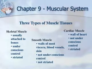
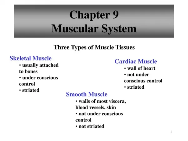
Chapter 6 Muscular System. Types of Muscles. Skeletal – striated & voluntary Smooth - involuntary Cardiac – heart Striated means it appears striped. Structure of a Muscle. Muscles are composed of many FIBERS that are arranged in bundles called FASCICLES.

Chapter 6 Muscular System
E N D
Presentation Transcript
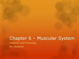
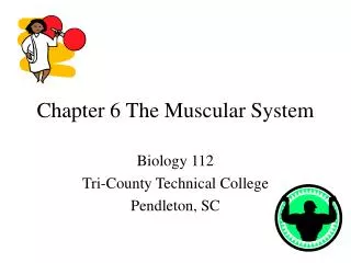
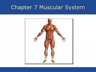
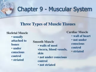
Types of Muscles • Skeletal – striated & voluntary • Smooth - involuntary • Cardiac – heart • Striated means it appears striped
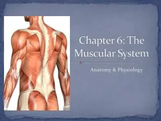
Structure of a Muscle • Muscles are composed of many FIBERS that are arranged in bundles called FASCICLES. • Muscles are separated from each other by FASCIA, which form the APONEUROSES and TENDONS that connect the muscles to bones. • Muscle Connective Tissue: • Epimysium – outermost layer that surrounds the entire muscle • Perimysium – separates and surrounds fascicles (bundles of muscle fibers) • Endomysium – surrounds each individual muscle fiber
Muscle Functions • The SOLE function of Muscular Tissue is to contract or shorten. As it contracts it: • Produces Movement • Maintains Posture • Stabilizes Joints • Generates Heat
Structure of a Muscle Fiber (muscle cells) • Each muscle cell is surrounded by a specialized cell membrane called the SARCOLEMMA. • The cytoplasm is called the SARCOPLASM. • Each muscle cell is filled with MYOFIBRILS which in turn are composed of MYOFILAMENTS.
Myofibril Structure • Myofibrils – contain myofilaments of myosin (thick) and actin (thin). • The myofilaments overlap to form I and A bands on the fiber: • DARK BANDS – A bands - myosin • LIGHT BANDS – I bands – actin • In the middle of each I band is a dark line called a Z line. • One Z line to the next is a SARCOMERE.
Neuromuscular Junction • The muscle fiber and the motor neuron make up the neuromuscular junction. • Motor end plate – folded area where the muscle and the neuron communicate • Synaptic cleft – the gap between the neuron and the motor end plate • Synaptic vesicles – where the neurotransmitters are stored
Neuromuscular Junction • The neurotransmitter that crosses the gap is ACETYLCHOLINE • ACH is broken down by CHOLINESTERASE.
Neuromuscular Junction • The theory of how a muscle contracts is the SLIDING FILAMENT THEORY. • The contraction occurs when the THICK filament (myosin) slides past the THIN filament (actin). • The sliding filament theory involves 5 different molecules plus CALCIUM • ATP • Myosin • Actin • Acetylcholine • Cholinesterase
Energy Source • The energy for muscle contraction comes from ATP provided by the process of CELLULAR RESPIRATION. • The molecule CREATINE PHOSPHATE helps with the regeneration of ATP. • Much of our body’s heat is produced by this process, which in turns helps maintain homeostasis.
Terms related to Muscle Contraction • Threshold stimulus • All or none response • Motor Unit • Recruitment • Muscle tone • Muscle Hypertrophy • Muscle Atrophy • Muscle fatigue • Muscle cramp • Oxygen debt • Origin and insertion • Tetanus • Rigor mortis
1. Threshold Stimulus Minimal strength to cause a contractionMotor neuron releases enough Acetylcholine to reach threshold 2. All-or-None Response Fibers don’t contract partially, they either do or they don’t