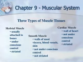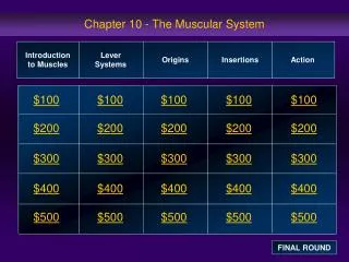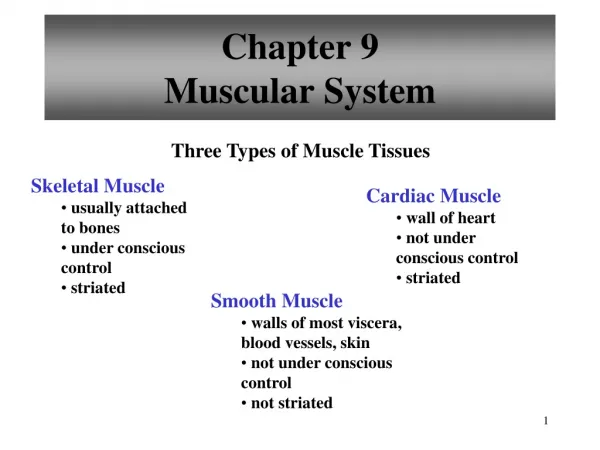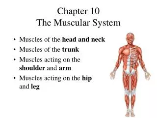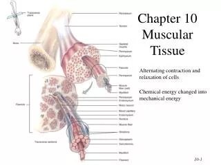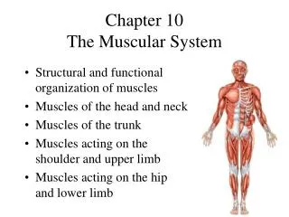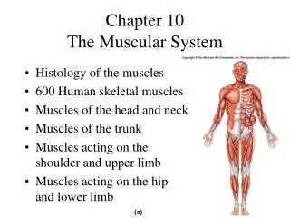
CHAPTER 10 MUSCULAR SYSTEM
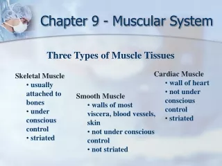
CHAPTER 10 MUSCULAR SYSTEM. 2 Muscles of the face & head (Aponeurosis/Fascia: Flat, sheet-like tendon) Figure 10.6 page: 331 A. Epicranius- frontalis & occipitalis 1. Word derivation a. Epi: (G) on top of b. Cranius: (L) skull 2. Origin: Occipital bone

CHAPTER 10 MUSCULAR SYSTEM
E N D
Presentation Transcript
CHAPTER 10 MUSCULAR SYSTEM
2 Muscles of the face & head • (Aponeurosis/Fascia: Flat, sheet-like tendon) • Figure 10.6 page: 331 • A. Epicranius- frontalis & occipitalis • 1. Word derivation • a. Epi: (G) on top of • b. Cranius: (L) skull • 2. Origin: Occipital bone • 3. Insertion: skin/muscles around eye • 4. Action: Raise eyebrows • B. Orbicularis Oculi • 1. Derivation: (L) circular/round & eye • 2. O: Maxilary & frontal bones • 3. I: Skin around eye • 4. A: Closes eye (Sphincter muscle)
4 C. Orbicularis Oris • 1. Oris (L) mouth • 2. O: Muscles near mouth • 3. I: skin of lips • 4. A: Closes and “pucker ups” lips (Sphincter) • D. Temporalis – video • 1. (L): side of head - temple region • temporalis bone • 2. O:Temporal bone • 3. I: Lateral surface of mandible • 4. A: Closes jaw #51 • E. Zygomaticus • 1. (G) Bar • 2. O: Zygomatic bone • 3. I: Obicularis oculi • 4. A: Raises corner of mouth (smile)
6 F. Buccinator – video #5 • 1. (L) Trumpeter • 2. O: Outer surface of maxilla & mandible • 3. I: Obicularis oris • 4. A: Compresses cheeks inwards • G. Masseter – video #31 • 1. (G) Chew • 2. O: Lower border of Zygomatic arch • 3. I: Lateral surface of mandible • 4. A: Closes jaw/chew
8 H. Sternocliedomastoid(eus)- SCM • 1. origins & insertions • 2. O: Sternum & clavicle(cliedeo, G-hook) • 3. I: mastoid process of temporal bone • 4. A: singly- rotate head, together- flex neck • I. Splenius capitis #47 • 1. • 2. O: Spinous & transverse processes of • lower cervical & upper thoracic vertebrae • 3. I: Mastoid process • 4. A: Rotate head, laterally flex neck
.11I. Semispinalis Capitis #42 1. semi = half (origin ½ & ½) 2. O: Spinous & transverse processes of lower cervical & upper thoracic vertebrae 3. I: Occipital bone 4. A: Extend, laterally flex & rotate neck
13 Muscles of the pectoral girdle • Posterior • Fig. 10.13 c; page 348 • A. Trapezius #58 • 1. Shape: trapezoid • 2. O: Occipital bone, spinous process of cervical & thoracic spine • 3. I: Clavicle; spine & acromion process of scapula 4. A: Raises & rotates scapula • B. Rhomboid Major #38 • 1. Shape rhomboid • 2. O: Spinous processes of upper thoracics • 3. I: Medial boarder of scapula • 4. A: Raises & Adducts scapula
15 C. Levator Scapulae #29 1. Lifts scapula 2. O: Transverse processes of C-spine 3. I: Margin(superior/medial) of scapula 4. A:raises & adducts scapula Pectoral girdle, anterior. Fig. 10.13 a pg. 347 D. Serratus anterior #44 1. (L) saw toothed 2. O: Outer surface of upper ribs 3. I: Ventral surface of scapula 4. A: Pulls scapula anterior & down (inferior)
18 E. Pectoralis minor #34Fig. 10.13 a; pg.347 1. (L) Chest 2. O: Sternal ends of upper ribs 3. I: Coracoid process (scapula) 4. A: Pulls scapula anterior & down Rotator cuff muscles(SITS) Fig. 10.14 b; page. 351 A. Supraspinatus* #50 1. Superior to scapula spine 2. O: Superior posterior aspect of scapula 3. I: Greater tubercle of humerous 4. A: Initiates arm abduction first 15 degrees
21 B. Infraspinatus #25 1. Inferior to scapular spine 2. O: Inferior posterior scapula 3. I: Greater tubercle of humerous 4. A: External rotation of arm C.Teres minor #53 1. (l) Long and round 2. O: Lateral boarder of scapula 3. Greater tubercle of humerous 4. A: Rotate arm externally
23 D. Subscapularis* #48Fig. 10.14 d; pg. 352 1. Underneath (anterior) side of scapular spine 2. O: Anterior surface of scapula 3. I: Lesser tubercle of humerous 4. A: Prime mover - Rotates arm internally
25 Muscles of the glenohumeral joint shoulder/upper arm Anterior muscles Figure 10.13 a; pg. 347 A. Pectoralis Major #33 1. (l) Chest 2. O: Clavicle, sternum & cartilagof upper ribs 3. I: Intertubercular grove of humerous 4. A: prime mover for upperar adduction
27 • B. Deltoid - #6 • figures 10.13 a&c pgs. 347 & 348 • Delta 1. (l) triangle shape • 2. O: Scapula, acromion process, spine, clavicle • 3. I: Deltoid tuberosity of humerous • 4. A: Prime mover for arm adduction (middle fibers), also flexes & extends arm
30 Posterior C. Latisimus dorsi (lats) #28 1. lats/wings 2. O: Spinous processes of sacral, lumbar & lower thoracic spine 3. I: Intertubercular grove 4. A: Prime mover for upper arm adduction Fig. 10.14b; pg. 251 Muscles that move the elbow Fig. 10.14 a&b pages: 351 & 352 Anterior chamber
32 A. Brachialis #3 1. (l) arm 2. Origin: anterior distal humerous 3. Insertion: coronoid process of ulna 4. Prime mover for forearm (elbow) flexion B. Biceps brachii #1 1. (l) arm & muscle has 2 heads 2. Origin: short head: coricoid process long head: glenoid tubercle & glenoidcavity 3. Insertion: Radial tuberosity 4. Not the prime mover- synergist for flexion; supination of wrist