Download
1 / 145
1.46k likes | 1.79k Vues
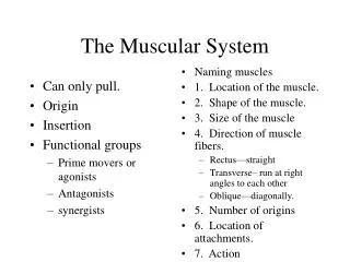
10 The Muscular System. Section 1: Functional Organization of the Muscular System. Learning Outcomes

E N D
10 The Muscular System
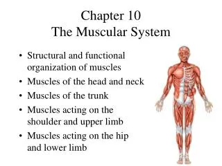
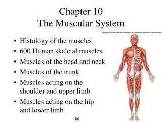
Section 1: Functional Organization of the Muscular System Learning Outcomes 10.1 Describe the arrangement of fascicles in the various types of muscles, explain the resulting functional differences, and explain how different classes of levers affect muscle efficiency. 10.2 Explain how the name of a muscle can help identify its location, appearance, or function. 10.3 Describe the separation of muscles into axial and appendicular divisions.
Section 1: Functional Organization of the Muscular System Muscular system Accounts for almost half body weight Contains ~700 muscles Vary widely in size, shape, and function Performance varies on how fibers are organized and how muscle attaches to skeleton Divided into two divisions Axial muscles Support and position axial skeleton Appendicular muscles Support, move, and brace the limbs
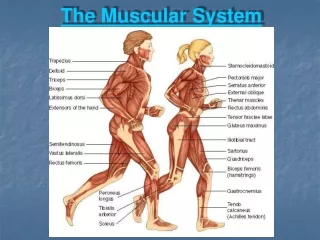
Figure 10 Section 1 The two divisions of the muscular system: axial muscles and appendicular muscles The proportion of total body weight contributed by the muscular system Axial muscles Lymphatic system 0.3% Urinary system 0.7% Reproductive system 0.15% Respiratory system 1.7% Endocrine system 0.15% Nervous system 2% Digestive system 6% Cardiovascular system 9% Skeletal system 20% Integumentary system 16% Muscular system 44% Appendicular muscles Tendons conduct the forces of contraction to perform specific tasks.
Module 10.1: Fascicle organization and range of motion Fascicle organization Parallel muscle Fascicles parallel to muscle long axis Most skeletal muscles in the body are parallel Some flat with broad attachments (aponeuroses) Some plump and cylindrical with tendon attachments Has central body (belly) Can contract until shortened by ~30% Due to muscle fiber shortening Example: biceps brachii
Figure 10.1 1 A parallel muscle: the biceps brachii (1) Fascicle Body (belly) 1
Module 10.1: Fascicle organization and range of motion Fascicle organization (continued) Convergent muscle Fascicles extending over broad area converge on common attachment site Versatile action Different portions can contract to produce different actions Entire muscle can contract Pulls less on attachment compared to parallel Example: pectoralis major
Figure 10.1 2 A convergent muscle: the pectoralis major (2) Base of muscle Tendon 2
Module 10.1: Fascicle organization and range of motion Fascicle organization (continued) Pennate muscle (penna, feather) Fascicles form common angle with tendon Pull at an angle on tendon Shorter movement of tendon versus parallel Contain more fibers than parallel of same size so produces more tension
Module 10.1: Fascicle organization and range of motion Fascicle organization (continued) Pennate muscle (continued) Three types Unipennate (all fibers on one side of tendon) Example: extensor digitorum Bipennate (fibers insert on both sides of tendon) Example: rectus femoris Multipennate (tendon branches within pennate muscle) Example: deltoid
Figure 10.1 3 Three pennate muscles Extensor digitorum muscle Rectus femoris muscle Deltoid muscle (3c) (3a) Tendons Extended tendon 3 If all the muscle fibers are on the same side of the tendon, the pennate muscle is unipennate. If a pennate muscle has fibers on both sides of the tendon, it is called bipennate. (3b) If the tendon branches within a pennate muscle, the muscle is said to be multipennate.
Figure 10.1 4 A circular muscle, or sphincter (4) 4 Contracted Relaxed
Module 10.1: Fascicle organization and range of motion Levers and Leverage Skeletal muscle force, speed, and direction depend on how it is attached to a lever Lever moves when applied force overcomes load Lever = rigid structure (bone) Fulcrum = fixed point on which lever pivots (joint) Applied force = muscle action Animation: Classes of Levers
Module 10.1: Fascicle organization and range of motion Levers and Leverage (continued) Lever classes First-class lever Fulcrum (F) between applied force (AF) and load (L) Acts like seesaw Balance depends on sizes of force and load and distribution on lever Few examples in body but important
Figure 10.1 5 A first-class lever Load Fulcrum Applied force
Module 10.1: Fascicle organization and range of motion Levers and Leverage (continued) Lever classes (continued) Second-class lever Load is between applied force and fulcrum Always farther from fulcrum than load so small force moves large load, but slowly and short distance Acts like wheelbarrow
Figure 10.1 6 A second-class lever Load Fulcrum Applied force
Module 10.1: Fascicle organization and range of motion Levers and Leverage (continued) Lever classes (continued) Third-class lever Most common lever in body Force applied between load and fulcrum Speed and distance traveled increased at expense of effective force
Figure 10.1 7 A third-class lever Applied force Load Fulcrum Biceps brachii muscle
Module 10.1 Review a. Define a lever, and describe the three classes of levers. b. The joint between the occipital bone of the skull and the first cervical vertebra (atlas) is which part of which class of lever system? c. Why does a pennate muscle generate more tension than does a parallel muscle of the same size?
Module 10.2: Muscle names Individual muscle parts Origin Fixed attachment Most are bones, some are connective tissue sheaths or bands (examples: intermuscular septa or interosseus membranes) Typically proximal to insertion in anatomical position Insertion Movable attachment Action Specific movement
Figure 10.2 1 The origins, insertion, and action of the biceps brachii muscle Origins of biceps brachii muscle Action Insertion of biceps brachii muscle
Module 10.2: Muscle names Muscles working together Agonist (prime mover) Muscle whose contraction chiefly responsible for producing particular movement Example: biceps brachii is agonist for elbow flexion Synergist (syn-, together + ergon, work) Muscle that helps larger agonist work efficiently May provide additional pull or stabilize origin Example: brachioradialis for elbow flexion
Module 10.2: Muscle names Muscles working together (continued) Antagonist Muscle whose action opposes particular agonist Example: triceps brachii for elbow flexion (to biceps brachii) Agonist for elbow extension
Figure 10.2 2 Descriptions of muscles based on their function Agonist, or prime mover Antagonist Synergist Origin of brachioradialis muscle Insertion of brachioradialis muscle
Module 10.2: Muscle names Muscle terminology examples Terms indicating specific body regions Abdominis (abdomen) Anconeus (elbow) Auricularis (auricle of ear) Brachialis (brachium) Terms indicating position, direction of fascicle organization Anterior (front) Externus (superficial) Extrinsic (outside) Inferioris (inferior)
Module 10.2: Muscle names Muscle terminology examples (continued) Terms indicating structural characteristics of muscle Name of origin Biceps (two heads) Triceps (three heads) Shape Deltoid (triangle) Orbucularis (circle) Other striking features Alba (white) Brevis (short)
Module 10.2: Muscle names Muscle terminology examples (continued) Terms indicating actions General Abductor Adductor Depressor Extensor Specific Buccinator (trumpeter) Risorius (laugher) Sartorius (like a tailor)
Module 10.2 Review a. Define the term synergist as it relates to muscle action. b. Muscle A abducts the humerus, and muscle B adducts the humerus. What is the relationship between these two muscles? c. What does the name flexor carpi radialis longus tell you about this muscle?
Module 10.3: Axial and appendicular divisions Axial muscles Arise on axial skeleton Encompass ~60% of skeletal muscles in body Position head and spinal column Move rib cage, assist breathing Appendicular muscles Stabilize or move appendicular skeleton Remaining 40% of all skeletal muscles
Module 10.3 Review a. What is the function of the axial muscles? b. Identify the division (axial or appendicular) to which each of the following muscles belongs: biceps brachii, external oblique, temporalis, and vastus medialis. c. Which structures labeled in the figures in this module are not muscles?
Section 2: Axial Muscles Learning Outcomes 10.4 Identify the principal muscles of facial expressions, along with their origins, insertions, and actions. 10.5Identify the principal muscles of the eye and jaw, along with their origins, insertions, and actions. 10.6Identify the principal muscles of the tongue, pharynx, and neck, along with their origins, insertions, and actions.
Section 2: Axial Muscles Learning Outcomes 10.7 Identify the principal muscles of the vertebral column, along with their origins, insertions, and actions. 10.8 Identify the principal muscles of the trunk, along with their origins, insertions, and actions. 10.9 Identify the principal muscles of the pelvic floor, along with their origins, insertions, and actions.
Section 2: Axial Muscles Axial Muscles Involved in stabilizing and positioning head, neck, and trunk Four groups Muscles of head and neck Muscles of vertebral column Muscles of thoracic and abdominal walls Muscles of pelvic floor
Figure 10 Section 2 The muscles of the head and neck that are not associated with the vertebral column The muscles of the vertebral column The muscles of the pelvic floor The oblique and rectus muscles of the trunk
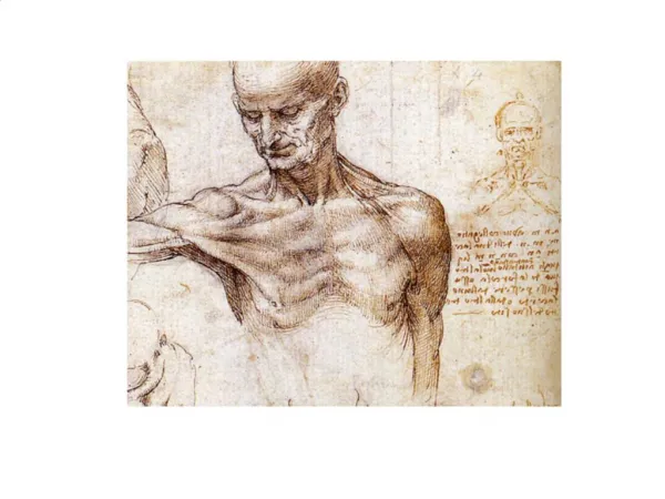
Module 10.4: Facial expression muscles Muscles of facial expression Originate on skull surface Insert into superficial fascia and dermis of skin Actions move skin A&P Flix: Buccinator Animation: Axial Muscles: Head and Neck
Figure 10.4 1 An anterior view showing superficial muscles on the right side of the face, and deeper muscles on the left side of the face. Epicranial aponeurosis Temporalis Frontal belly of occipitofrontalis Corrugator supercilii Orbicularis oculi Procerus Nasalis Levator labii superioris Zygomaticus minor Levator anguli oris Zygomaticus major Masseter Orbicularis oris Buccinator Risorius Depressor anguli oris Platysma Depressor labil inferioris Mentalis (cut) Thyroid cartilage of the larynx Clavicle
Figure 10.4 2 A lateral view showing the major facial muscles Frontal belly of the occipitofrontalis muscle Occipital belly of the occipito- frontalis muscle Epicranial aponeurosis Orbicularis oculi Nasalis Muscles of the Mouth and Cheek Levator labii superioris Zygomaticus minor Zygomaticus major Buccinator Orbicularis oris Risorius Mentalis (cut) Depressor labil inferioris Temporalis Depressor anguli oris Masseter
Module 10.4 Review a. Identify the muscles associated with the mouth. b. State whether the following muscles involve the mouth, eye, nose, ear, scalp, or neck: buccinator, corrugator supercilii, mentalis, nasalis, platysma, procerus, and risorius. c. Explain how an individual is able to consciously move the skin on the scalp but is not able to consciously move the skin of the thigh.
Module 10.5: Muscles of external eye and mastication Extrinsic eye muscles position the eye Six muscles insert on surface of eye and originate in orbit Muscles of mastication move the lower jaw All mastication muscles are seen on the lateral sides of the skull
Figure 10.5 1 – 2 A lateral view of the right eye (left) showing five extrinsic eye muscles, and a medial view of the right eye (right) showing the sixth extrinsic eye muscle Superior rectus Frontal bone Superior rectus Superior oblique Superior oblique Levator palpebrae superioris Optic nerve Trochlea (ligamentous sling) Inferior oblique Inferior rectus Lateral rectus Optic nerve Medial rectus Maxilla
Module 10.5: Muscles of external eye and mastication A&P Flix: Temporalis A&P Flix: Masseter
Figure 10.5 3 – 4 Two anterior views of the right eye: the directions of eye movements produced by each extrinsic eye muscle operation independently (left), and the origins of the extrinsic eye muscles in the orbit (right) Levator palpebrae superioris Trochlear nerve (IV) Superior rectus Trochlea Superior oblique Trochlea Superior rectus Superior oblique Oculomotor nerve (III) Medial rectus Lateral rectus Lateral rectus Medial rectus Abducens nerve (VI) Optic nerve (II) Inferior oblique Inferior rectus Inferior oblique Inferior rectus
Figure 10.5 6 A superficial lateral view showing the largest muscles of mastication of the right side of the head Superior temporal line Temporalis Capsule of temporomandibular joint Zygomatic arch Masseter
Figure 10.5 7 A lateral view showing the pterygoid muscles after removal of the superficial muscles and the right mandibular ramus Lateral pterygoid Medial pterygoid Cut edge of mandible
Module 10.5 Review a. Name the extrinsic eye muscles. b. Which muscles have their origin on the lateral pterygoid plates and their insertion on the medial surface of the mandibular ramus? c. If you were contracting and relaxing your masseter muscle, what would you probably be doing?
Module 10.6: Muscles of the tongue, pharynx, and neck Muscles of the tongue are closely associated with pharynx and neck muscles Actions of these muscles assist in speaking and chewing Many help support the hyoid bone
Figure 10.6 1 – 2 A lateral view showing the tongue muscles as dissected after removal of the left half of the mandible Styloid process Palatoglossus Styloglossus Genioglossus Hyoglossus Hyoid bone Mandible (cut)