Download
1 / 23
230 likes | 255 Vues
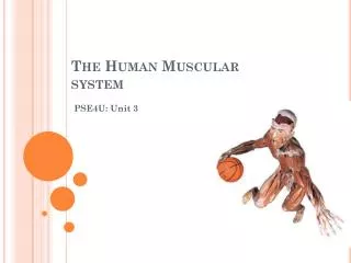
Explore the functions, types, and structure of the muscular system, understanding how muscles work to facilitate movement, maintain posture, generate heat, and enable communication. Discover the characteristics of skeletal muscles and the intricate organization of muscle fibers. Dive into the details of skeletal muscle cells and their membrane potentials. Uncover the neuromuscular communication process and how muscle contraction is initiated through the release of acetylcholine.

E N D
Introduction • Most abundant tissue in the body • Always working • Movement • Locomotion • Breathing • Sitting/standing upright • Beating heart
Muscular System Functions • Body movement • Via muscle contraction; create overall body movements • Posture maintenance • Constant skeletal muscle tone • Respiration • Thoracic muscles • Body heat production • Heat released as byproduct of skeletal muscles • Communication • Oral, written, body language • Organ & vessel contraction • Move/mix food & water in GI tract, excrete secretions, regulate blood flow • Heart beat • Cardiac muscle contraction; propels blood to body
Types of Muscle • Skeletal muscle • Cardiac muscle • Smooth muscle
Characteristics of Skeletal Muscles • ~40% BW • Attached to skeleton • Contractility • Muscles can shorten with force causing movement of structures to which they’re attached; lengthen passively (gravity or force of opposing muscle) • Excitability • Muscles respond to stimuli (usually nerves cause contraction of skeletal muscle) • Extensibility • Skeletal muscle can be stretched to resting length and a little beyond • Elasticity • Ability of muscles to recoil to original resting length after stretching
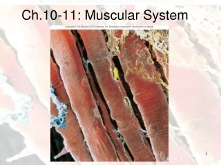
Structure of Skeletal Muscle • Hierarchical organization: • Each muscle (organ) surrounded by epimysium (upon + muscle) /fascia (fillet) – connective tissue • Fasciculus (bundle) – bundles of muscle cells/fibers; surrounded by perimysium (around + muscle) – loose connective tissue • Several muscle fibers per fasciculus; surrounded by endomyosium (within + muscle) – loose connective tissue
Structure of Skeletal Muscle Cells • Microscopic structure • Each fiber composed of myofibrils • Myofibrils composed of myofilaments (protein filaments) arranged in sarcomeres (flesh + part) • Actin myofilaments (thin) • Myosin myofilaments (thick) • Each muscle fiber has: • Sarcolemma (cell membrane) • Sarcoplasm (cytoplasm) • Sarcroplasmic reticulum (ER) - ↑[Ca2+] – muscle contractions • Transverse tubules (T tubules) – network of tubes connecting sarcolemma to sarcoplasmic reticulum
Structure of Skeletal Muscle Cells • Banding Pattern (striations) (See 7.3 a) • I-band (I-LIGHT): thin myofilaments only (actin) • A-band (A-DARK): thick & thin myofilaments (myosin & actin) • Z-disk: dark line in middle of I-band; site of actin attachment • H-zone: lighter, central region in middle of A-band; thick myofilaments only (myosin) • M-line: dark line in middle of H-zone; site of myosin attachment • Sarcomere: functional contractile unit of muscle cell; runs from A-disk to Z-disk
Membrane Potentials • Outside of most cell membranes is positively charged relative to inside • A resting membrane potential is created = charge difference across the membrane of a resting cell • Occurs because: • Higher [K+] inside than outside • Cell membrane more permeable to K+ than other ions (K+ channels open) • Action potential = change in membrane potential in an excitable tissue that is propagated as an electrical signal • Made via Sodium-Potassium pump.
Resting membrane potential. Na+ channels & some K+ channels closed. A few K+ diffuse down c.g. through open K+ channels, making outside positive. • Depolarization. Na+ channels open. A few Na+ diffuse down c.g. through open Na+ channels, making inside positive. • Repolarization. Na+ channels closed; Na+ movement into cell stops. K+ channels open. K+ movement outside cell increases, making outside positive.
Neuromuscular Communication • Motor neurons innervate all muscle tissue • The “motor unit”: 1 motor neuron + all muscle fibers it stimulates • From 2-3 fibers/unit (fine control) to 2000 fibers/unit (large power movements) • Neuromuscular junction (NMJ) structures: • Presynaptic terminals – bulbous ends of motor neuron (ends of axons) • NMJ or synapse – region of chemical communication between cells • Synaptic vesicles contain neurotransmitter (acetylcholine) • Synaptic cleft – space between presynaptic terminals and postsynaptic membrane (sarcolemma)
Neuromuscular Communication, continued.. • Basic events of muscle contraction: • Action potential (nerve impulse) arrives at presynaptic terminals • Synaptic vesicles release acetylcholine (ACh) into synaptic cleft via exocytosis • ACh binds to receptors sites on Na+ channels in postsynaptic membrane, causing them to open • Na+ moves into cell causing an action potential which travels along sarcolemma & T-tubules • ACh triggers Ca2+ release from sarcoplasmic reticulum • Ca2+ inside muscle triggers actin & myosin “sliding filament” action • ATP is spent • Acetylcholinesterase degrades remaining ACh to limit contraction stimulus
Neuromuscular Communication, continued.. • Sliding Filament Model • Actin & myosin myofilaments slide past one another causing sarcomeres to shorten • H-zones and I-bands shorten; A-bands maintain constant width (Fig 7.7)
Neuromuscular Communication, continued.. • Sliding Filament Model, continued… • Ca2+ binds to troponin • Tropomysin molecules slide into groove • Myosin attaches to exposed sites on actin myofilament • Energy from ATP stored in myosin heads • Myosin heads bind to actin forming cross-bridges • Stored energy used to myosin heads causing actin myofilament to slide • ATP binds to myosin causing head to release & return to resting position • Cycle repeats if Ca2+ still bound to troponin & ATP still available
Muscle Twitch, Summation, Tetanus, & Recruitment • Muscle twitch - contraction of a muscle fiber in response to a stimulus • Lag phase – time between stimulus application and beginning of contraction • Contraction phase – time of contraction • Relaxation phase – time of relaxation
Muscle Twitch, Summation, Tetanus, & Recruitment, continued… • Strength of Muscle Contraction (↑’d by:) • Summation – increasing force of contraction of muscle fibers in muscle • Recruitment – increasing number of muscle fibers contracting within muscle • Tetanus (convulsive tension) – sustained muscular contraction caused by a series of nerve stimuli repeated so rapidly that there is no relaxation, instead a sustained contraction results
Energy Requirements for Muscle Contraction • Mitochondria produce ATP • Creatine phosphate is energy storage • Used to generate ATP • Anaerobic respiration (w/o O2) • Generates 2 ATP; short-lived • Lactic acid as waste product (irritant) • Aerobic respiration (w/ O2) • Generates up to 38 ATP; long-term • Uses other nutrient molecules (F.A., a.a.) • Oxygen debt – must be repaid after labor
Fatigue • Psychological fatigue – CNS causes the perception that continued muscle contraction is impossible • Muscle fatigue – force of muscle contraction becomes increasingly weak when ATP is used faster than can be produced and lactic acid builds up faster than it can be removed • Physiological contracture – muscles cannot contract or relax (not enough ATP to do either)
Types of Muscle Contractions • Isometric contractions (equal distance) – tension increases, muscle same length • Isotonic contractions (equal tension) – tension is constant, muscle shortens • Concentric – tension increases as muscle shortens • Eccentric – tension constant as muscle lengthens