Download
1 / 36
360 likes | 564 Vues
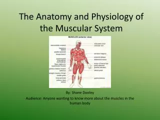
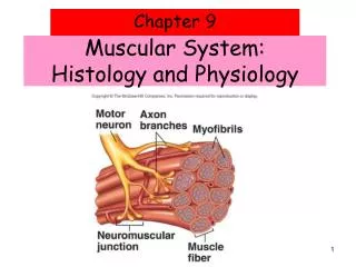
Muscular System: Histology and Physiology. Muscular System Functions. Body movement (Locomotion) Maintenance of posture Respiration Diaphragm and intercostal contractions Communication (Verbal and Facial) Constriction of organs and vessels Peristalsis of intestinal tract

E N D
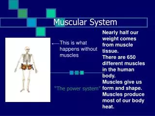
Muscular System Functions • Body movement (Locomotion) • Maintenance of posture • Respiration • Diaphragm and intercostal contractions • Communication (Verbal and Facial) • Constriction of organs and vessels • Peristalsis of intestinal tract • Vasoconstriction of b.v. and other structures • Heart beat • Production of body heat
Properties of Muscle • Excitability: capacity of muscle to respond to a stimulus • Contractility: ability of a muscle to shorten and generate pulling force • Extensibility: muscle can be stretched back to its original length • Elasticity: ability of muscle to recoil to original resting length after stretched
Types of Muscle • Skeletal • Attached to bones • Makes up 40% of body weight • Responsible for locomotion, facial expressions, posture, respiratory movements, other types of body movement • Voluntary in action; controlled by somatic motor neurons • Smooth • In the walls of hollow organs, blood vessels, eye, glands, uterus, skin • Some functions: propel urine, mix food in digestive tract, dilating/constricting pupils, regulating blood flow, • In some locations, autorhythmic • Controlled involuntarily by endocrine and autonomic nervous systems • Cardiac • Heart: major source of movement of blood • Autorhythmic • Controlled involuntarily by endocrine and autonomic nervous systems
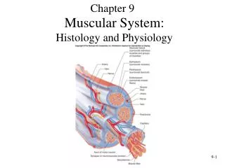
Connective Tissue Sheaths • Connective Tissue of a Muscle • Epimysium. Dense regular c.t. surrounding entire muscle • Separates muscle from surrounding tissues and organs • Connected to the deep fascia • Perimysium. Collagen and elastic fibers surrounding a group of muscle fibers called a fascicle • Contains b.v and nerves • Endomysium. Loose connective tissue that surrounds individual muscle fibers • Also contains b.v., nerves • Collagen fibers of all 3 layers come together at each end of muscle to form a tendon.
Nerve and Blood Vessel Supply • Motor neurons • stimulate muscle fibers to contract • Neuron axons branch so that each muscle fiber (muscle cell) is innervated • Form a neuromuscular junction (= myoneural junction) • Capillary beds surround muscle fibers • Muscles require large amts of energy • Extensive vascular network delivers necessary oxygen and nutrients and carries away metabolic waste produced by muscle fibers
Basic Features of a Skeletal Muscle • Muscle attachments • Most skeletal muscles run from one bone to another • One bone will move – other bone remains fixed • Origin – less movable attach- ment • Insertion – more movable attach- ment
Basic Features of a Skeletal Muscle • Muscle attachments (continued) • Muscles attach to origins and insertions by connective tissue • Fleshy attachments – connective tissue fibers are short • Indirect attachments – connective tissue forms a tendon.
Skeletal Muscle Structure • Composed of muscle cells (fibers), connective tissue, blood vessels, nerves • Fibers are long, cylindrical, and multinucleated • Tend to be smaller diameter in small muscles and larger in large muscles. 1 mm- 4 cm in length • Develop from myoblasts • Striated appearance • Nuclei are peripherally located
Muscle Fiber Anatomy • Sarcolemma - cell membrane • Surrounds the sarcoplasm(cytoplasm of fiber) • Contains many of the same organelles seen in other cells • An abundance of the oxygen-binding protein myoglobin • Punctuated by openings called the transverse tubules (T-tubules) • Narrow tubes that extend into the sarcoplasm at right angles to the surface • Filled with extracellular fluid • Myofibrils -cylindrical structures within muscle fiber • Are bundles of protein filaments (=myofilaments) • Two types of myofilaments • Actin filaments (thin filaments) • Myosin filaments (thick filaments) • At each end of the fiber, myofibrils are anchored to the inner surface of the sarcolemma • When myofibril shortens, muscle shortens (contracts)
Sarcoplasmic Reticulum (SR) • SR is an elaborate, smooth endoplasmic reticulum • runs longitudinally and surrounds each myofibril • Form chambers called terminal cisternae on either side of the T-tubules • A single T-tubule and the 2 terminal cisternae form a triad • SR stores Ca++ when muscle not contracting • When stimulated, calcium released into sarcoplasm • SR membrane has Ca++ pumps that function to pump Ca++ out of the sarcoplasm back into the SR after contraction
Sarcoplasmic Reticulum (SR) Figure 9.5
Sarcomeres: Z Disk to Z Disk • Sarcomere - repeating functional units of a myofibril • About 10,000 sarcomeres per myofibril, end to end • Each is about 2 µm long • Differences in size, density, and distribution of thick and thin filaments gives the muscle fiber a banded or striated appearance. • A bands: a dark band; full length of thick (myosin) filament • M line - protein to which myosins attach • H zone - thick but NO thin filaments • I bands: a light band; from Z disks to ends of thick filaments • Thin but NO thick filaments • Extends from A band of one sarcomere to A band of the next sarcomere • Z disk: filamentous network of protein. Serves as attachment for actin myofilaments • Titin filaments: elastic chains of amino acids; keep thick and thin filaments in proper alignment
Myosin (Thick) Myofilament • Many elongated myosin molecules shaped like golf clubs. • Single filament contains roughly 300 myosin molecules • Molecule consists of two heavy myosin molecules wound together to form a rod portion lying parallel to the myosin myofilament and two heads that extend laterally. • Myosin heads • Can bind to active sites on the actin molecules to form cross-bridges. (Actin binding site) • Attached to the rod portion by a hinge region that can bend and straighten during contraction. • Have ATPase activity: activity that breaks down adenosine triphosphate (ATP), releasing energy. Part of the energy is used to bend the hinge region of the myosin molecule during contraction
Actin (Thin) Myofilaments • Thin Filament: composed of 3 major proteins • F (fibrous) actin • Tropomyosin • Troponin • Two strands of fibrous (F) actin form a double helix extending the length of the myofilament; attached at either end at sarcomere. • Composed of G actin monomers each of which has a myosin-binding site (see yellow dot) • Actin site can bind myosin during muscle contraction. • Tropomyosin: an elongated protein winds along the groove of the F actin double helix. • Troponin is composed of three subunits: • Tn-A : binds to actin • Tn-T :binds to tropomyosin, • Tn-C :binds to calcium ions.
Sliding Filament Model of Contraction • Thin filaments slide past the thick ones so that the actin and myosin filaments overlap to a greater degree • In the relaxed state, thin and thick filaments overlap only slightly • Upon stimulation, myosin heads bind to actin and sliding begins
Sliding Filament Model of Contraction • Each myosin head binds and detaches several times during contraction, acting like a ratchet to generate tension and propel the thin filaments to the center of the sarcomere • As this event occurs throughout the sarcomeres, the muscle shortens PLAY InterActive Physiology®: Muscular System: Sliding Filament Theory
Neuromuscular Junction • Region where the motor neuron stimulates the muscle fiber • The neuromuscular junction is formed by : 1. End of motor neuron axon (axon terminal) • Terminals have small membranous sacs (synaptic vesicles) that contain the neurotransmitter acetylcholine(ACh) 2. The motor end plate of a muscle • A specific part of the sarcolemma that contains ACh receptors • Though exceedingly close, axonal ends and muscle fibers are always separated by a space called the synaptic cleft
Neuromuscular Junction Figure 9.7 (a-c)
Motor Unit: The Nerve-Muscle Functional Unit • A motor unit is a motor neuron and all the muscle fibers it supplies • The number of muscle fibers per motor unit can vary from a few (4-6) to hundreds (1200-1500) • Muscles that control fine movements (fingers, eyes) have small motor units • Large weight-bearing muscles (thighs, hips) have large motor units
Motor Unit: The Nerve-Muscle Functional Unit Figure 9.12 (a)
Motor Unit: The Nerve-Muscle Functional Unit • Muscle fibers from a motor unit are spread throughout the muscle • Not confined to one fascicle • Therefore, contraction of a single motor unit causes weak contraction of the entire muscle • Stronger and stronger contractions of a muscle require more and more motor units being stimulated (recruited)
Smooth Muscle • Cells are not striated • Fibers smaller than those in skeletal muscle • Spindle-shaped; single, central nucleus • More actin than myosin • No sarcomeres • Not arranged as symmetrically as in skeletal muscle, thus NO striations. • Caveolae: indentations in sarcolemma; • May act like T tubules • Dense bodies instead of Z disks • Have noncontractile intermediate filaments
Smooth Muscle • Grouped into sheets in walls of hollow organs • Longitudinal layer – muscle fibers run parallel to organ’s long axis • Circular layer – muscle fibers run around circumference of the organ • Both layers participate in peristalsis Figure 9.24
Smooth Muscle • Is innervated by autonomic nervous system (ANS) • Visceral or unitary smooth muscle • Only a few muscle fibers innervated in each group • Impulse spreads through gap junctions • Who sheet contracts as a unit • Often autorhythmic • Multiunit: • Cells or groups of cells act as independent units • Arrector pili of skin and iris of eye
Cardiac Muscle • Found only in heart where it forms a thick layer called the myocardium • Striated fibers that branch • Each cell usually has one centrally-located nucleus • Fibers joined by intercalated disks • IDs are composites of desmosomes and gap junctions • Allow excitation in one fiber to spread quickly to adjoining fibers • Under control of the ANS (involuntary) and endocrine system (hormones) • Some cells are autorhythmic • Fibers spontaneously contract (aka Pacemaker cells)
Cardiac Muscle Tissue Figure 10.10a
Disorders of Muscle Tissue • Muscle tissues experience few disorders • Heart muscle is the exception • Skeletal muscle – remarkably resistant to infection • Smooth muscle – problems stem from external irritants
Disorders of Muscle Tissue • Muscular dystrophy – a group of inherited muscle destroying disease • Affected muscles enlarge with fat and connective tissue • Muscles degenerate • Types of muscular dystrophy • Duchenne muscular dystrophy • Myotonic dystrophy
Disorders of Muscle Tissue • Myofascial pain syndrome – pain is caused by tightened bands of muscle fibers • Fibromyalgia – a mysterious chronic-pain syndrome • Affects mostly women • Symptoms – fatigue, sleep abnormalities, severe musculoskeletal pain, and headache
Developmental Aspects: Regeneration • Cardiac and skeletal muscle become amitotic, but can lengthen and thicken • Myoblast-like satellite cells show very limited regenerative ability • Cardiac cells lack satellite cells • Smooth muscle has good regenerative ability • There is a biological basis for greater strength in men than in women • Women’s skeletal muscle makes up 36% of their body mass • Men’s skeletal muscle makes up 42% of their body mass
Developmental Aspects: Male and Female • These differences are due primarily to the male sex hormone testosterone • With more muscle mass, men are generally stronger than women • Body strength per unit muscle mass, however, is the same in both sexes
Developmental Aspects: Age Related • With age, connective tissue increases and muscle fibers decrease • Muscles become stringier and more sinewy • By age 80, 50% of muscle mass is lost (sarcopenia) • Decreased density of capillaries in muscle • Reduced stamina • Increased recovery time • Regular exercise reverses sarcopenia