Download
1 / 19
190 likes | 478 Vues
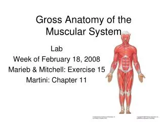
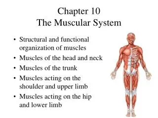
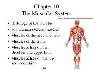
Chapter 10 Anatomy of the Muscular System. Introduction (Figures 10-5 and 10-6). There are more than 600 skeletal muscles in the body From 40% to 50% of body weight is skeletal muscle Muscles, along with the skeleton, determine the form and contour of the body.

E N D
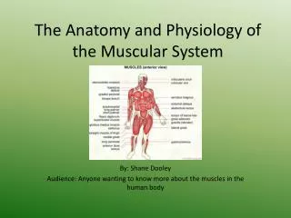
Introduction (Figures 10-5 and 10-6) • There are more than 600 skeletal muscles in the body • From 40% to 50% of body weight is skeletal muscle • Muscles, along with the skeleton, determine the form and contour of the body
Skeletal Muscle Structure (Figure 10-1) • Connective tissue components • Endomysium—delicate connective tissue membrane that covers specialized skeletal muscle fibers • Perimysium—tough connective tissue binding together fascicles • Epimysium—coarse sheath covering the muscle as a whole • These three fibrous components may become a tendon or an aponeurosis
Skeletal Muscle Structure • Size, shape, and fiber arrangement (Figure 10-2) • Skeletal muscles vary considerably in size, shape, and fiber arrangement • Size—range from extremely small to large masses • Shape—variety of shapes such as broad, narrow, long, tapering, short, blunt, triangular, quadrilateral, irregular, flat sheets, or bulky masses • Arrangement—variety of arrangements such as parallel to long axis, converge to a narrow attachment, oblique, pennate, bipennate, or curved; the direction of fibers is significant because of its relationship to function
Skeletal Muscle Structure • Attachment of muscles (Figure 10-3) • Origin—point of attachment that does not move when muscle contracts • Insertion—point of attachment that moves when muscle contracts
Skeletal Muscle Structure • Muscle actions • Most movements are produced by the coordinated action of several muscles; some muscles in the group contract while others relax • Prime movers (agonists)—muscles or groups of muscles that directly perform a specific movement • Antagonists—muscles that, when contracting, directly oppose prime movers; relax while prime movers (agonists) are contracting to produce movement; provide precision and control during contraction of prime movers • Synergists—muscles that contract at the same time as the prime movers; they facilitate prime movers’ actions to produce a more efficient movement • Fixator muscles—joint stabilizers
Skeletal Muscle Structure • Lever systems • In the human body, bones serve as levers and joints serve as fulcrums; contracting muscle applies a pulling force on a bone lever at the point of the muscle’s attachment to the bone, causing the insertion bone to move about its joint-fulcrum • Lever system—composed of four component parts (Figure 10-4): • Rigid bar (bone) • Fulcrum (F) around which the rod moves (joint) • Load (L) that is moved • Pull (P) that produces movement (muscle contraction)
Skeletal Muscle Structure • Lever systems (cont.) • First-class levers • Fulcrum lies between the pull and the load • Not abundant in human body; serve as levers of stability • Second-class levers • Load lies between the fulcrum and the joint at which pull is exerted • Controversy exists regarding presence of these levers in the human body • Third-class levers • Pull is exerted between the fulcrum and the load • Permit rapid and extensive movement • Most common type of lever found in the body
How Muscles Are Named • Muscle names can be in Latin or English (this book uses English) • Muscles are named using one or more of the following features: • Location, function, or shape (Tables 10-1; 10-2; 10-3) • Direction of fibers—named according to fiber orientation (Table 10-4) • Number of heads or divisions (Table 10-4) • Points of attachment—origin and insertion points • Relative size—small, medium, or large (Table 10-5) • Names supply hints on how to deduce muscle actions
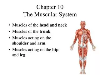
Important Skeletal Muscles • Muscles of facial expression—unique in that at least one point of attachment is to the deep layers of the skin over the face or neck (Figures 10-7 and 10-8; Table 10-6) • Muscles of mastication—responsible for chewing movements (Figure 10-9; Table 10-6) • Muscles that move the head—paired muscles on either side of the neck are responsible for head movements (Figure 10-10; Table 10-7)
Trunk Muscles • Muscles of the thorax—critical importance in respiration (Figure 10-9; Table 10-8) • Muscles of the abdominal wall—arranged in three layers, with fibers in each layer running in different directions to increase strength (Figure 10-12; Table 10-9) • Muscles of the back—bend or stabilize the back (Figure 10-13; Table 10-10) • Muscles of the pelvic floor—support the structures in the pelvic cavity (Figure 10-14; Table 10-11)
Upper Limb Muscles • Muscles acting on the shoulder girdle—muscles that attach the upper extremity to the torso are located anteriorly (chest) or posteriorly (back and neck); these muscles also allow extensive movement (Figure 10-15; Table 10-12) • Muscles that move the upper arm—the shoulder is a synovial joint allowing extensive movement in every plane of motion (Figure 10-17; Table 10-13)
Upper Limb Muscles • Muscles that move the forearm—found proximally to the elbow and attach to the ulna and radius (Figures 10-19 and 10-20; Table 10-14) • Muscles that move the wrist, hand, and fingers—these muscles are located on the anterior or posterior surfaces of the forearm (Figures 10-21 through 10-23; Table 10-15)
Lower Limb Muscles • The pelvic girdle and lower extremity function in locomotion and maintenance of stability • Muscles that move the thigh and lower leg (Figures 10-5, 10-6, and 10-24 through 10-30; Tables 10-16 and 10-17)
Lower Limb Muscles • Muscles that move the ankle and foot (Figures 10-31 and 10-32; Table 10-18) • Extrinsic foot muscles are located in the leg and exert their actions by pulling on tendons that insert on bones in the ankle and foot; responsible for dorsiflexion, plantar flexion, inversion, and eversion • Intrinsic foot muscles are located within the foot; responsible for flexion, extension, abduction, and adduction of the toes
Posture • Maintaining the posture of the body is one of the major roles muscles play • “Good posture”—body alignment that most favors function and requires the least muscular work to maintain, keeping the body’s center of gravity over its base
Posture • How posture is maintained • Muscles exert a continual pull on bones in the opposite direction from gravity • Structures and systems other than muscle and bones have a role in maintaining posture • Nervous system—responsible for determining muscle tone and also regulation and coordination of the amount of pull exerted by individual muscles • Respiratory, digestive, excretory, and endocrine systems all contribute to maintain posture
Cycle of Life: Muscular System • Muscle cells—increase or decrease in number, size, and ability to shorten at different periods • Pathological conditions at different periods may affect the muscular system
Cycle of Life: Muscular System • Life cycle changes—manifested in other components of functional unit • Infancy and childhood—coordination and controlling of muscle contraction permits sequential development steps • Degenerative changes of advancing age result in replacement of muscle cells with nonfunctional connective tissue • Diminished strength