Download
1 / 45
460 likes | 539 Vues
Explore the structure and functions of skeletal muscles, including unique characteristics, tissue composition, and neuromuscular junctions. Learn about muscle fiber structure, sarcomere organization, and types of contractions. Understand the classification, naming conventions, and sizes of skeletal muscles. Illustrated with over 200 detailed images. Ideal resource for medical students, healthcare professionals, and anatomy enthusiasts.

E N D
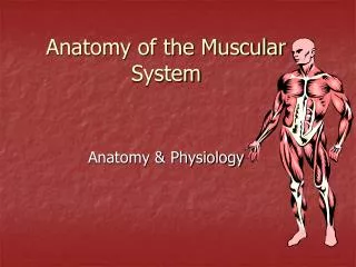
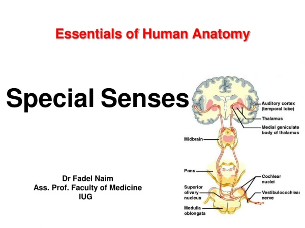
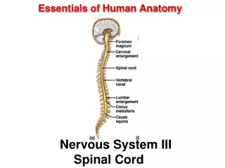
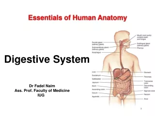
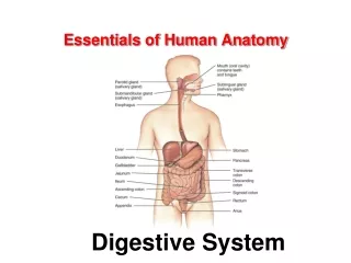
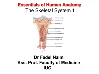
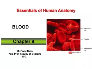
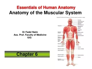
Essentials of Human Anatomy Anatomy of the Muscular System Chapter 6 Dr Fadel Naim Ass. Prof. Faculty of Medicine IUG
Introduction • There are more than 600 skeletal muscles in the body • From 40% to 50% of body weight is skeletal muscle • Muscles, along with the skeleton, determine the form and contour of the body
Functions of skeletal Muscles • Body movements • Maintaining Posture • Stabilizing Joints • Body positions • Generating heat
4 Unique Characteristics of Muscle Tissue • Excitability is equated with responsiveness. • Contractility causes the fiber to shorten resulting in either a pull on bones or the movement of specific body parts. • Elasticity is the muscle’s ability to return to its original length when tension is released. • Extensibility is capability of extending in length in response to the contraction of opposing muscle fibers.
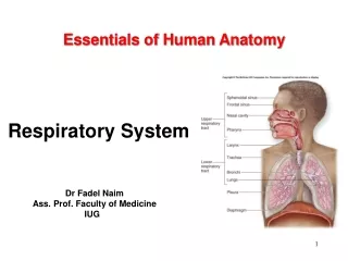
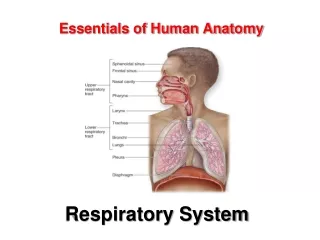
Structure of a Skeletal Muscle • Skeletal Muscle • organ of the muscular system • - skeletal muscle tissue • - nervous tissue • - blood • - connective tissues • fascia • tendons • aponeuroses
Skeletal Muscle Tissue • Skeletal muscles are organs • Vary in shape and size • A skeletal muscle is composed of cells • Each cell is as long as the muscle • Small muscle: 100 micrometers long; 10 micrometers in diameter • Large muscle: 35 centimeters long; 100 micrometers in diameter
Skeletal Muscle Structure • Connective tissue components • Endomysium—delicate connective tissue membrane that covers specialized skeletal muscle fibers • Perimysium—tough connective tissue binding together fascicles • Epimysium—coarse sheath covering the muscle as a whole • These three fibrous components may become a tendon or an aponeurosis
Muscle Fiber Structure • Multiple nuclei • Sarcolemma • T-tubules • Sarcoplasmic reticulum • Sarcoplasm • Mitochondria • Glycogen & ions • Myofibrils
Skeletal Muscle Has Striations • Appearance is due to size and density differences between thick filaments and thin filaments. • Under the light microscope, two differently shaded bands are present. • The dark bands, called A bands, contain the entire thick filament. • At either end of a thick filament is a region where thin filaments extend into the A band between the stacked thick filaments. • Light bands, called I bands, contain thin filaments only. • I band is lighter shaded than an A band because only the thin filaments occupy this region.
Sarcomere: Organization of Fibers • Z disks • I band • A band • H zone • M line
Neuromuscular Junction • also known as myoneural junction • site where an axon and muscle fiber meet • motor neuron • motor end plate • synapse • synaptic cleft • synaptic vesicles • neurotransmitters
Motor Unit • single motor neuron • all muscle fibers controlled by motor neuron
skeletal muscles Classification • By the way fascicles are organized • By relationships of fascicles to tendons • 4 patterns of fascicle organization: • parallel • convergent • pennate • circular
Parallel Muscles • Fibers parallel to the long axis of muscle • e.g., biceps brachii • The center or body of the muscle thickens when parallel muscle contracts • Parallel muscles contract about 30%
Convergent Muscles • A broad area converges on attachment site (tendon, aponeurosis, or raphe) • Muscle fibers pull in different directions, depending on stimulation • e.g., pectoralis muscles
Pennate Muscles • Unipennate: • fibers on 1 side of tendon e.g., extensor digitorum • Bipennate: • fibers on both sides of tendon e.g., rectus femoris • Multipennate: • tendon branches within muscle e.g., deltoid
Circular Muscles • Also called sphincters • Open and close to guard entrances of body • e.g., obicularis oris
Types of Contractions • isotonic – muscle contracts and changes length • concentric – shortening contraction • isometric – muscle contracts but does not change length • eccentric – lengthening contraction
Descriptive Names or Skeletal Muscles • Location in the body-identifies body regions: • e.g., temporalis muscle • Origin and insertion-First part of name indicates origin, second part of name indicates insertion: e.g., genioglossus muscle • Fascicle organization-Describes fascicle orientation within muscle: i.e., rectus (straight), transversus, oblique • Relative position- • Externus (superficialis):visible at body surface • Internus (profundus):deep muscles • Extrinsic:muscles outside an organ • Intrinsic:muscles inside an organ • Structural characteristics • Number of tendons: bi = 2, tri = 3 • Shape: trapezius, deltoid, rhomboid • Size • Action
Names for Muscle Size • Major = larger • Maximus = largest • Minor = small • Minimus = smallest • Longus = long • Longissimus = longest • Teres = long and round • Brevis = short • Magnus = large • Action • Movements: • e.g., flexor, extensor, retractor
Muscle Atrophy • Reduction in muscle size, tone, and power. • Due to reduced stimulation, it loses both mass and tone. • Muscle becomes flaccid, and its fibers decrease in size and become weaker. • Even a temporary reduction in muscle use can lead to muscular atrophy.
Muscle Hypertrophy • An increase in muscle fiber size. • Muscle size may be improved by exercising. • Repetitive, exhaustive stimulation of muscle fibers results in more mitochondria, larger glycogen reserves, and an increased ability to produce ATP. • Ultimately, each muscle fiber develops more myofibrils, and each myofibril contains a larger number of myofilaments.
Three Types of Skeletal Muscle Fibers • Fast • are large in diameter • contain large glycogen reserves • densely packed myofibrils • relatively few mitochondria • called white fibers due to lack of myoglobin • majority of skeletal muscle fibers in the body • Intermediate • resemble fast fibers; however • have a greater resistance to fatigue • Slow • smaller and they • contract more slowly • called red fibers because due to myoglobin
Posture • Maintaining the posture of the body is one of the major roles muscles play • “Good posture”—body alignment that most favors function and requires the least muscular work to maintain, keeping the body’s center of gravity over its base
Posture • How posture is maintained • Muscles exert a continual pull on bones in the opposite direction from gravity • Structures and systems other than muscle and bones have a role in maintaining posture • Nervous system—responsible for determining muscle tone and also regulation and coordination of the amount of pull exerted by individual muscles • Respiratory, digestive, excretory, and endocrine systems all contribute to maintain posture
Cycle of Life: Muscular System • Life cycle changes—manifested in other components of functional unit • Infancy and childhood—coordination and controlling of muscle contraction permits sequential development steps • Degenerative changes of advancing age result in replacement of muscle cells with nonfunctional connective tissue • Diminished strength
Cycle of Life: Muscular System • Muscle cells—increase or decrease in number, size, and ability to shorten at different periods • Pathological conditions at different periods may affect the muscular system
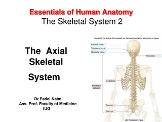
Axial Muscles • Have both their origins and insertions on parts of the axial skeleton. • Support and move the head and spinal column. • Function in nonverbal communication by affecting facial features. • Move the lower jaw during chewing. • Assist in food processing and swallowing. • Aid breathing. • Support and protect the abdominal and pelvic organs. • Are not responsible for stabilizing or moving the pectoral or pelvic girdles or their attached limbs.
Appendicular Muscles • Control the movements of the upper and lower limbs. • Stabilize and control the movements of the pectoral and pelvic girdles. • Organized into groups based on their location in the body or the part of the skeleton they move. • Work in groups that are either synergistic or antagonistic.
Appendicular Muscles • Organized into specific groups. • muscles that move the pectoral girdle • muscles that move the glenohumeral joint/arm • arm and forearm muscles that move the elbow joint/forearm • forearm muscles that move the wrist joint, hand, and fingers • intrinsic muscles of the hand
Intramuscular Injections • The gluteus maximus is a large, thick muscle with coarse Fasciculi that can be easily separated without damage. • The great thickness of this muscle makes it ideal for intramuscular Injections. • To avoid injury to the underlying Sciatic nerve, the injection should be given well forward On the upper outer quadrant of the buttock.