Chapter 10 Muscular Tissue
660 likes | 1.17k Vues
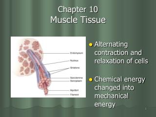
Chapter 10 Muscular Tissue. Lecture slides prepared by Curtis DeFriez, Weber State University. Functions of Muscular Tissue. Like nervous tissue, muscles are excitable or "irritable” they have the ability to respond to a stimulus Unlike nerves, however, muscles are also:

Chapter 10 Muscular Tissue
E N D
Presentation Transcript
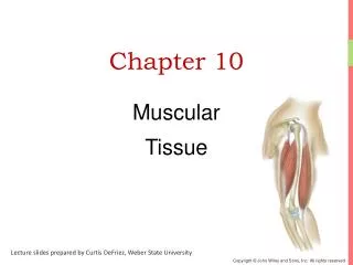
Chapter 10 Muscular Tissue Lecture slides prepared by Curtis DeFriez, Weber State University
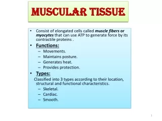
Functions of Muscular Tissue • Like nervous tissue, muscles are excitable or "irritable” • they have the ability to respond to a stimulus • Unlike nerves, however, muscles are also: • Contractible(they can shorten in length) • Extensible(they can extend or stretch) • Elastic(they can return to their original shape)
Functions of Muscular Tissue • Muscle makes up a large percentage of the body’s weight • Their main functions are to: • Create motion– muscles work with nerves, bones, and joints to produce body movements • Stabilize body positions and maintain posture • Store substances within the body using sphincters • Move substances by peristaltic contractions • Generate heat through thermogenesis
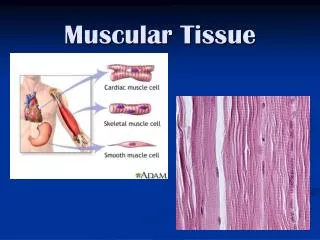
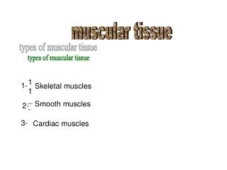
Three Types of Muscular Tissue (a) Skeletal muscle (c) Visceral smooth muscle (b) Cardiac muscle
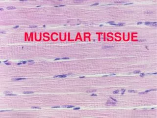
Skeletal Muscle • Skeletal muscle fibers are very long “cells” - next to neurons (which can be over a meter long), perhaps the longest in the body • The Sartorious muscle contains single fibers that are at least 30 cm long A single skeletal muscle fiber
Skeletal Muscle The terminal processes of a motor neuron in close proximity to the sarcolemma of a skeletal muscle fiber Motor neuron Sarcolemma
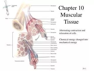
Organization of Muscle Tissue • The epimysium, perimysium, and endomysium all are continuous with the connective tissues that form tendons and ligaments (attach skeletal muscle to bone) and muscle fascia (connect muscles to other muscles to form groups of muscles)
Organization of Muscle Tissue Epimysium Perimysium Organization of a single muscle belly
Organization of Muscle Tissue Organization of a fasciculus
Organization of Muscle Tissue Organization of a muscle fiber
Organization of Muscle Tissue A muscle, a fasciculus, and a fiber all visualized
Organization of Muscle Tissue • In groups of muscles the epimysium continues to become thicker, forming fascia which covers many muscles • This graphic shows the fascia lata enveloping the entire group of quadriceps and hamstring muscles in the thing
Organization of Muscle Tissue • Many large muscle groups are encased in both a superficial and a deep fascia Real Anatomy, John Wiley and Sons
Organization of Muscle Tissue Epicranial aponeurosis • An aponeurosis is essentially a thick fascia that connects two muscle bellies. This epicranial aponeurosis connects the muscle bellies of the occipitalis and the frontalis to form “one” muscle: The occipitofrontalis Frontal belly of the occipitofrontalis m.
Organization of Muscle Tissue Veins, arteries, and nerves are located in the deep fascia between muscles of the thigh.
The Skeletal Muscle Fiber • Beneath the connective tissue endomysium is found the plasma membrane (called the sarcolemma) of an individual skeletal muscle fiber • The cytoplasm (sarcoplasm) of skeletal muscle fibers is chocked full of contractile proteins arranged in myofibrils
The Skeletal Muscle Fiber • You should learn the names of the internal structures of the muscle fiber • Sarcolemma • Sarcoplasm • Myofibril • T-tubules • Triad (with terminal cisterns • Sarcoplasmic reticulum • Sarcomere
The Skeletal Muscle Fiber • Increasing the level of magnification, the myofibrils are seen to be composed of filaments • Thick filaments • Thin filaments
The Skeletal Muscle Fiber • The basic functional unit of skeletal muscle fibers is the sarcomere: An arrangement of thick and thin filaments sandwiched between two Z discs A scanning electron micrograph of a sarcomere
The Skeletal Muscle Fiber • Muscle contraction occurs in the sarcomeres The “Z line” is really a Z disc when considered in 3 dimensions. A sarcomere extends from Z disc to Z disc.
Muscle Proteins • Myofibrils are built from three groups of proteins • Contractile proteins generate force during contraction • Regulatory proteins help switch the contraction process on and off • Structural proteins keep the thick and thin filaments in proper alignment and link the myofibrils to the sarcolemma and extracellular matrix
Muscle Proteins • The thin filaments are comprised mostly of the structural protein actin, and the thick filaments are comprised mostly of the structural protein myosin • However, in both types of filaments, there are also other structural and regulatory proteins
Muscle Proteins • In the thin filaments actin proteins are strung together like a bead of pearls • In the thick filaments myosin proteins look like golf clubs bound together
Muscle Proteins • In this first graphic, the myosin binding sites on the actin proteins are readily visible. • The regulatory proteins troponin and tropomyosin have been added to the bottom graphic: The myosin binding sites have been covered
Muscle Proteins • In this graphic the troponin-tropomyosin complex has slid down into the “gutters” of the actin molecule unblocking the myosin binding site • The troponin-tropomyosin complex can slide back and forth depending on the presence of Ca2+ Myosin binding site exposed
Muscle Proteins • Ca2+ binds to troponin which changes the shape of the troponin-tropomyosin complex and uncovers the myosin binding sites on actin
Muscle Proteins • Besides contractile and regulatory proteins, muscle contains about a dozen structural proteins which contribute to the alignment, stability, elasticity, and extensibility of myofibrils • Titan is the third most plentiful protein in muscle, after actin and myosin - it extends from the Z disc and accounts for much of the elasticity of myofibrils • Dystrophin is discussed later as it relates to the disease of muscular dystrophy
The Sliding-Filament Mechanism • With exposure of the myosin binding sites on actin (the thin filaments)—in the presence of Ca2+ and ATP—the thick and thin filaments “slide” on one another and the sarcomere is shortened
The Sliding-Filament Mechanism • The “sliding” of actin on myosin (thick filaments on thin filaments) can be broken down into a 4 step process
Step 1: ATP hydrolysis • Step 2: Attachment
Step 3: Power Stroke • Step 4: Detachment
Contraction and Movement OverviewInteractions Animation • Contraction and Movement You must be connected to the internet to run this animation.
Excitation-Contraction Coupling • We will come back to the term excitation-contraction coupling in a little bit • Before we can describe the entire process, from thinking of moving a muscle to actual contraction of sarcomeres, we must first explore the processes that occur at the neuromuscular junction
Neuromuscular Junction • Excitation-Contraction coupling (EC coupling) involves events at the junction between a motor neuron and a skeletal muscle fiber
Neuromuscular Junction • An enlarged view of the neuromuscular junction • The presynaptic membrane is on the neuron while the postsynaptic membrane is the motor end plate on the muscle cell. The two membranes are separated by a space, or “cleft”
Neuromuscular Junction • Conscious thought (to move a muscle) results in activation of a motor neuron, and release of the neurotransmitter acetylcholine (AcCh) at the NM junction • The enzyme acetylcholinesterase breaks down AcCh after a short period of time
Neuromuscular Junction • The plasma membrane on the “far side” of the NMJ belongs to the muscle cell and is called the motor end plate • The motor end plate is rich in chemical (ligand) - gated sodium channels that respond to AcCh. Another way to say this: The receptors for AcCh are on the ligand-gated sodium channels on the motor end plate
Neuromuscular Junction • The chemical events at the NMJ transmit the electrical events of a neuronal action potential into the electrical events of a muscle action potential
Neuromuscular JunctionInteractions Animation • Neuromuscular Junctions You must be connected to the internet to run this animation.
Muscle Action Potential • The muscle AP is propagated over the surface of the muscle cell membrane (sarcolemma) via voltage (electrical)-gated Na+ and K+ channels
Generating An Action Potential • The flow of ions through cell a membrane looks a lot like a "piece" of electricity flowing through a wire (but not as fast) • Generating an AP on the muscle membrane involves the transfer of information from an electrical signal (down the neuron), to a chemicalsignal (at the NMJ), back to an electrical signal (depolarization of the sarcolemma) • This added complexity (changing from electrical to chemical back to electrical signals) provides necessary control of the process
Sources of Muscle Energy • Stored ATP • 3 seconds • Energy transferred from stored creatine phosphate • 12 seconds • Aerobic ATP production • Anaerobic glucose use • 30-40 seconds
Skeletal Muscle Metabolism • In a state of homeostasis, muscle use of O2 and nutrients is balanced by the production of manageable levels of waste products like • CO2 • Heat - 70-80% of the energy used by muscles is lost as heat - muscle activity is important for maintaining body temperature • Lactic acid (anaerobic)
Skeletal Muscle Metabolism • Oxygen Debt, or "Excess Post-Exercise Oxygen Consumption" (EPOC) is the amount of O2 repayment required after exercise in skeletal muscle to: • Replenish ATP stores • Replenish creatine phosphate and myoglobin stores • Convert lactic acid back into pyruvate so it can be used in the Krebs cycle to replenish ATP