MUSCULAR TISSUE
MUSCULAR TISSUE. Objectives: By the end of this lecture you should be able to: Identify and describe the histological structure of the three types of muscle cells and list the differences between them. MUSCULAR TISSUE. Made of elongated muscle cells ( fibers ).

MUSCULAR TISSUE
E N D
Presentation Transcript
MUSCULAR TISSUE Objectives: By the end of this lecture you should be able to: • Identify and describe the histological structure of the three types of muscle cells and list the differences between them.
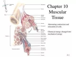
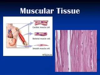
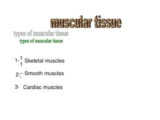
MUSCULAR TISSUE • Made of elongated muscle cells (fibers). • 3 types of muscles (muscle fibers): 1- Skeletal: striated, voluntary. 2- Cardiac: striated, involuntary. 3- Smooth: non-striated, involuntary.
SKELETAL MUSCLE • The whole muscle is covered by a C.T. covering, the epimysium. • Consists of parallel skeletal muscle fibers, arranged in bundles, separated by C.T. septa, the perimysium. • The individual fibers are separated by C.T., endomysium,
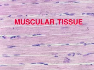
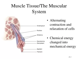
Skeletal Muscle Fibers • L.M. Picture: • Cylindrical in shape. • Non-branched. • Covered by a clear cell membrane, the sarcolemma. • Multinucleated: nuclei are multiple and are peripherally located (close to the sarcolemma). • Cytoplasm(sarcoplasm)is acidophilic and shows clear transverse striations.
Skeletal Muscle Fibers • E.M. Picture: Sarcoplasm contains: • Parallel myofibrils. • Numerous mitochondria, arranged in rows between the myofibrils. • Well developed smooth endoplasmic reticulum (sarcoplasmic reticulum). • Myoglobin pigment. • Glycogen.
Skeletal Muscle Fibers • E.M. Picture of Myofibrils: • Contractile threads (organelles), arranged longitudinally in the sarcoplasm. • Each myofibril shows alternating dark (A) and light bands (I). • The A band shows a pale area in the middle (H band) which is divided by a dark line (M line). • The (I) band shows a dark line in the middle (Z line).
Skeletal Muscle Fibers • E.M. Picture of Myofibrils: • The sarcomere is the distance between 2 successive Z lines. • The myofibrils are formed of myofilaments (thick myosin and thin actin). • The (A) band is formed of myosinmyofilaments mainly and the terminal ends of actin myofilaments. • The (I) band is formed of actinmyofilaments. Sarcomere Z Z I I A A A A A A Myosin Actin
Skeletal Muscle Fibers • The TRIAD tubular system: • The sarcolemma sends transverse invaginations into the sarcoplasm, the T-tubules. They form collars around the myofibrils at the level of theA - I junctions. • The SR forms transverse wider cisternae (terminal cisternae) on either side of the T-tubule. • The 2 terminal cisternae of the SR and the T-tubule in-between them form the triad tubular system, which plays an important role during muscle contraction.
CARDIAC MUSCLE • Found in the myocardium. • Striated and involuntary. • L.M. Picture of Cardiac Muscle Fibers: • Cylindrical in shape. • Intermediate in diameter between skeletal and smooth muscle fibers. • Branch and anastomose. • Covered by a thin sarcolemma. • Mononucleated. Nuclei are oval and central. • Sarcoplasm shows non-clear striations (fewer myofibrils). • Divided into short segments (cells) by the intercalated discs.
Cardiac Muscle Fibers • E.M. Picture: • Few myofibrils. • Numerous mitochondria. • Less abundant SR. • T-tubules come in contact with only one cisterna of SR forming “Diads” (not triads). • Glycogen & myoglobin. • Intercalated discs: are formed of the two cell membranes of 2 successive cardiac muscle cells, connected together by junctional complexes (desmosomes and gap junctions).
SMOOTH MUSCLE • Present in walls of blood vessels and viscera (digestive, urinary, genital .... etc). • Non-striated and involuntary. • L.M. Picture of Smooth Muscle Fibers: • Fusiform in shape (spindle-shaped). • Small diameter. • Non-branched. • Thin sarcolemma. • Mononucleated. Nuclei are oval & central in position. • Sarcoplasm is non-striated.
Smooth Muscle Fibers • E.M. Picture: • Sarcoplasm contains mitochondria and sarcoplasmic reticulum. • Myosin & actinfilaments are irregularly arranged (that’s why no striations could be observed). • Cells are connected together by gap junctions for cell communication.
REGENERATION OF MUSCLE (1) Skeletal muscle cells: - Can not divide. - Limited regeneration by satellite cells (stem cells on the muscle cell’s surface). (2) Cardiac muscle cells: -No regenerative capacity. (3) Smooth muscle cells: - Can divide. - Regenerate from pericytes. → active regenerative response.
Comparison between different types of muscle fibers SKELETAL CARDIAC SMOOTH Site Muscle attached to skeleton Myocardium of the heart Viscera, e.g. stomach Shape Cylindrical Cylindrical Fusiform Diameter Largest Medium-sized Smallest Branching Non-branched Branched Non-branched Striations Clear Not clear Absent Intercalated discs Absent Present Absent Nuclei Numerous and peripheral One central nucleus One central nucleus Action Voluntary Involuntary Involuntary Regeneration Limited No Yes
Clinical Application Cardiac hypertrophy: • During cardiac hypertrophy the number of cardiac muscle cells is not increased; instead, they become longer and larger in diameter.