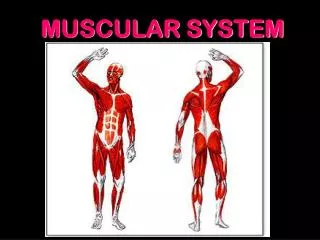
Muscular tissue :
Muscular tissue : Muscular tissue & hoeostasis: muscular tissue contribute to homeostasi by producing body movements , moving substances through the body & producing heat to mentain normal body temperature.

Muscular tissue :
E N D
Presentation Transcript
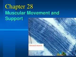
Muscular tissue : Muscular tissue & hoeostasis: muscular tissue contribute to homeostasi by producing body movements , moving substances through the body & producing heat to mentain normal body temperature. Motion result from alternating contruction &relaxation of musclerswhich form about 40 -50% of total body weight .muscular strength reflect the primary function of muscle (trnasformation of chemical energy to mechanical energy to generate force , perform work , produce movemernt ., in addition muscle stasbilize body position regulate organ volume , generate heat , propel fluid & food matter through various body system , scientfic study of muscle known as myology. Types of muscular tissue : 1- skeletal m.t.: it is straited (alternating dark &light protein bands), it work in voluntary manner . 2- cardiac m.t.:muscle of heart , also straited , but involuntary , the heart has a pacemaker that intiates contraction this termed autorhythmicity. 3– smooth m.t.located in the wall of hollow organ like blood vessels, airways,most of abdominal &pelvic organs. It is not straited , &involuntary .
Function of muscular tissue : 1- producing body movement 2- stbilizing body position : skeletal m.contraction stabilize joints &help mentain body position . 3- storing &mving substances within the body: storage is accomplished by sustained contraction of ringlike bands of smooth m.called sphincters which prevent outflow of contents of hollow organ , cardiac m. cont.pump blood through blood vessels of body , cont.&relaxation of blood vessels help adjust blood v. diameter &thus regulate the rate of blood flow. Smooth m. cont. also move food &substances like bile &enzymes through GIT. 4– generating heat :as muscular t. contract , it produce heat ,this calledv thermogenesis.Properties of muscular tissue : 1-electrical excitability : both nerve & muscle have ability to respond to certain stimuliby producing electrical signals called action potentials (impulses) , two masin type of stimuli bring action potential one is autorhythmic in m.t. itself as in heart ., other is chemical such as neurotransmitters, hormones. 2- contractility:is the abillity of m. to contract when stimulated by an action potential. 3-extendsibility:is the ability of m.t. to stretch , whin limit without being damaged 4- elasticity:is the ability of m,t. to return to its original length& shape after cont &extension .
Skeletal m.t.: sk.m. composed of 100 or 1000s of cells called m. fiber or m.cell. Sk.m contain connective t. surrounding m. fiber. Connective t. components: 3 types of con. T.: The outermost layer encircling the entire m.( epimysium). Perimysium it surrounds groups of 10 – 100 or more m. fiber , seperating them into bundle called fasicles Endomysium penetrate the interior of each fasicle& seperates individual m.fiber from one another , all 3 connective t.layers may extend beyond the m. f.to form (tendon) which attach m. to bone microscopic anatomy of sk.m.fiber: Most important componant of s.m.f. are m.fiber , the diameter of which 10- 100 microm., the typical length of s.m.f. is 10 cm. The multiple nuclei of s.m.f. located just beneath the sarcolemma( the plasma membrane of m. f.). Tiny of invaginations of the sarcol.,called transverse T- tubules, turned in from the surface toward the center of each m. fiber , because they open to the outside of fiber they are fillef with interstitial fluid . M. action potential tavel along the sarcolema& through T-tubule , quickley spread through out the m. fiber .this arrangement ensure that an action p. excite all parts of m. f. at same instant . Within sarcolema is the sarcoplasm (cytoplasm of m.f.) it include substantial amount of glycogen can used for synthesis of ATP also contain protein called myoglobin (this portein found only in m. binds oxygen that diffuse in to m.f. from interstitial fluid , myoglobin release oxygen when it needed by mitocondria for ATP production .
Muscular tissue : Muscular tissue & hoeostasis: muscular tissue contribute to homeostasi by producing body movements , moving substances through the body & producing heat to mentain normal body temperature. Motion result from alternating contruction &relaxation of musclerswhich form about 40 -50% of total body weight .muscular strength reflect the primary function of muscle (trnasformation of chemical energy to mechanical energy to generate force , perform work , produce movemernt ., in addition muscle stasbilize body position regulate organ volume , generate heat , propel fluid & food matter through various body system , scientfic study of muscle known as myology. Types of muscular tissue : 1- skeletal m.t.: it is straited (alternating dark &light protein bands), it work in voluntary manner . 2- cardiac m.t.:muscle of heart , also straited , but involuntary , the heart has a pacemaker that intiates contraction this termed autorhythmicity. 3– smooth m.t.located in the wall of hollow organ like blood vessels, airways,most of abdominal &pelvic organs. It is not straited , &involuntary .
Skeletal Muscle Structure the muscle is covered in a layer of connective tissue known as the Epimysium. The Epimysium protects the muscle from friction against other muscles and bones.
It also continues at the end of the muscle to form (along with other connective tissues) the muscles tendon. at the cross section of the muscle you can see bundles of fibres, known as Fasciculi, which are surrounded by another connective tissue, called the Perimysium. Each Fasciculi contains anywhere between 10 and 100 muscle fibres, depending on the muscle in question. A large strong muscle, such as thoses forming Quadriceps would have a large number of fibres within each bundle. A smaller muscle used for precision movement, such as those in the hand would contain far fewer fibres per Fasciculi.
Looking at each muscle fibre in detail, you can see they too are covered in a fibrous connective tissue, known as Endomysium which insulates each muscle fibre. Muscle fibres can range from 10 to 80 micrometers in diameter and may be up to 35cm long. Beneath the Endomysium and surrounding the muscle fibre is the Sarcolemma which is the fibres cell membrane and beneath this is the Sarcoplasm, which is the cells cytoplasm, a gelatinous fluid which fills most cells. This contains Glycogen and Fats for energy and also Mitochondria which are the cells power houses, inside which the cells energy is produced.
Each muscle fibre itself contains cylindrical organelles known as Myofibrils. Each muscle fibre contains hundreds to thousands of Myofibrils. Each myofibril has Actin and Myosin filaments. which run the length of the muscle fibre and are important in muscle contraction. The myosin & actin filaments partially interdigitate & thus cause the myofibrile to have alternate light &dark bands the light band contain only actin filament &called I bandbecause they are isotropic to polarised light ,the dark band contain myosin filament &are called A band because they are anisotropic to polarised light ,,,
also ther are small projections from the side of myosin filament ,these are cross bridge ,they project entire the long of the filament except in the center ,interaction between cross bridge & actin filament causes contruction.the end of actin filaments are attached to a so called • Z- disc, from this disc filaments extend in both direction to interdigitate with myosin filament , the portion of myofibrile that lies between 2 successive z discs called sarcomere
Surrounding the Myofibril there is a network of tubules and channels called the Sarcoplasmic Reticulum in which Calcium is stored which is important in muscle contraction., The SARCOLEMMA has a unique feature: it has holes in it. These "holes" lead into tubes called TRANSVERSE TUBULES (or T-TUBULES for short). These tubules pass down into the muscle cell and go around the MYOFIBRILS. However, these tubules DO NOT open into the interior of the muscle cell; they pass completely through and open somewhere else on the sarcolemma (i.e., these tubules are not used to get things into and out of the muscle cell). The function of T-TUBULES is to conduct impulses from the surface of the cell (SARCOLEMMA) down into the cell and, specifically, to the SARCOPLASMIC RETICULUM.
Myosin filament: Each myosin filament composed of multiple Myosin molecules , each molecule compesd of 6 polypeptide chain 2 heavy chain & 4 light chains, the 2 heavy chain wrap spirally arroun each other forming double helix , at the end each form globular structure called head , the light chain are part of the head , 2 light chain at each head.. The double helix are called the taile of myosin molecule.
Part of each helix portion of myosin molecule extends to the side along with head , forming the cross bridges. The head of myosin filament has ATPase activity to provide energy for muscle contraction .
Actin filament : It composed of 3 components: actin, tropomyosin &troponin , the actin filament is a double strands F-actin protein molecule , each is composed of polymerised G- actin molecule for each one actin molecule is attached to one molecule of ADP which is the active site on the actin with which the cross bridge of myosin filament attached to cause muscle cotraction.
The other molecule is tropomyosin these molecule are connected loosely with F-actin strands, the 3rd molecule is troponin which composed of 3 subunits : Troponin I which has strong affinity for actin Trponin T for tropomyosin Troponin C for ca+
The active sites of actin inhibeted in relaxed muscle by the troponin- tropomyosin complex & Ca+ inhibit the inhibitory effect of the complex. . Since sarcomeres are arranged in series, each adds to the shortening of the whole muscle. Stronger muscles have many cells arranged in parallel like the jaw muscle. Fast, but less powerful muscles, like the triceps, are composed of fewer longer fibers. .
General mechanism of muscle contruction : The muscle contruction occure in the fallowing steps : 1- action potential travels along amotor nerve to its end on muscle fiber. 2- at each ending the nerve secrets amount of neurotransmitter called acetylcholine. 3- the acetylcholine act on alocal area of the muscle fiber membrane to open multiple Ach- gated chanels through protein molecule in the muscle fiber membrane.
4-Openning of Ach channels allows large quantities of Na+ ion to flow to the interior of the muscle fiber membrane at the point of nerve terminal this intiate action potential in the muscle fiber . 5-action potential travels along the muscle fiber membrane. 6- the action potential depolarizes the muscle fiber membrane & travels deeply where it cause the sarcoplasmic reticulum to release alarge quantities of Ca+ .
7- the Ca+ ion intiate attractive force between actin & myosin filaments causing them to slide together which is the conrtuctile process:Myosin head energized via myosin-ATPase activity which converts the bound ATP to ADP + Pi 8 - Calcium binds to troponin 9 - Tropomyosin translocates to uncover the cross-bridge binding sites 10- The energized myosin binding sites approach the binding sites
11- The first myosin head binds to actin 12 - The bound myosin head releases ADP + Pi, flips and the muscle shortens 13 - The second myosin head binds to actin 14 - The first myosin head binds ATP to allow the actin and myosin to unbind 15 - The second myosin head releases its ADP + Pi, flips & the muscle shortens further 16 - The second myosin head binds to ATP to allow the actin and myosin to unbind
17 - The second myosin head unbinds from the actin, flips back and is ready for the next cycle 18 - The cross-bridge cycle is terminated by the loss of calcium from the troponin 19 - Tropomyosin translocates to cover the cross-bridge binding sites 20 - The calcium returns to the sarcoplasmic reticulum, the muscle relaxes & returns to the resting state untle new action potential comes along
Skeletal muscles contract according to the sliding filament model: An action potential originating in the CNS reaches an alpha motor neuron, which then transmits an action potential down its own axon. The action potential propagates by activating sodium dependent channels along the axon toward the synaptic cleft. Eventually, the action potential reaches the motor neuron terminal and causes a calcium ion influx through the calcium-dependent channels.
3-The Ca2+ influx causes vesicles containing the neurotransmitter acetylcholine to fuse with the plasma membrane, releasing acetylcholine out into the extracellular space between the motor neuron terminal and the motor end plate of the skeletal muscle fiber. 4 -The acetylcholine diffuses across the synapse and binds to and activates nicotinic acetylcholine receptors on the motor end plate of the muscle cell. Activation of the nicotinic receptor opens its intrinsic sodium/potassium channel, causing sodium to rush in and potassium to trickle out. Because the channel is more permeable to sodium, the muscle fiber membrane becomes more positively charged, triggering an action potential
5- The action potential spreads through the muscle fiber's network of T-tubules, depolarizing the inner portion of the muscle fiber. 6- The depolarization activates calcium channels in the T tubule membrane, which are in close proximity to calcium-release channels in the adjacent sarcoplasmic reticulum. 7 - Activated voltage-gated calcium channels physically interact with calcium-release channels to activate them, causing the sarcoplasmic reticulum to release calcium
8-The calcium binds to the troponin C present on the actin-containing thin filaments of the myofibrils. The troponin then modulates the tropomyosin. Normally the tropomyosin obstructs binding sites for myosin on the thin filament; once calcium binds to the troponin C and causes an change in the troponin protein, troponin T allows tropomyosin to move, unblocking the binding sites.
9-Myosin (which has ADP and inorganic phosphate ) binds to the newly uncovered binding sites on the thin filament (binding to the thin filament is very tightly coupled to the release of inorganic phosphate). Myosin is now bound to actin in the strong binding state. The release of ADP and inorganic phosphate are tightly coupled to the power stroke (actin acts as a cofactor in the release of inorganic phosphate,). This will pull the Z-bands towards each other, thus shortening the sarcomere and the I-band. 10- ATP binds myosin, allowing it to release actin and be in the weak binding state (a lack of ATP makes this step impossible).
Mechanical Properties of Muscle Types of muscle contraction: In an isotonic contraction, the muscle + tendon length shortens, as would happen if one lifted a heavy ball. If a limb moves a lot, the muscle fibers can shorten a great deal and become more inefficient. An isometric contraction is one in which the muscle + tendon length remains the same. In other words, the muscle can only shorten as much as the tendon stretches. This would happen if you were trying to lift a 1000 pound table top.
Excitatition – contraction coupling : An increase in Ca+concentration in the sarcoplasm starts muscle contraction & decrease stope it . When the muscle relaxed the concentration of Ca+ is very low . Huge amount of ca+is stored inside the S.R.as muscle action potential propagates along the sarcolemma &into the T tubules , it cause ca+ release channel in the SR to open , when these channel open ,ca+ flows out of the SRinto the sarcoplasm around the thick &thin filament , the released ca+ combine with troponin, causing it to change in shape , this conformational changes moves trpomyosin away from myosine binding sites once these binding sites are free ,myosin heades bind to them to form cross bridge & contraction cycle begins . The SR also contain ca+ active transport pump that use ATP to move ca+ constantly from the sarcplasm into the SR , While m. action potentials propagate through the T-tubule ,the ca+ release channel are open , after the last A.P has propagated throughout T- tubules ca+ release channels close as the opump moves ca+ back into the SR, the concentration of ca+ in the sarcolplasm decrease inside SR ca+ binding protein (calsequestrin) bind ca+ enablin\g more ca+ to be stored in SR.
Length –tension relationship: The forcefulness of m.contraction depends on the length of sarcomers ,within the m. before cont. begins,at a sarcomers length (2-2.4micro m.), the zone of overlap in each sarcomere is optimal & the m.f. can develop maximum tension (100%) which occure when the zone of overlap between thick &thin filament extends from the edge of H-zone (the center of A-band where only thick filament & no thin filament )to the end of thick filament . As the sarcomeres stretched , the zone of overlap shortens& fewer myosin head can make contact with thin filament , therefore , the tension the fiber produce decrease , when s.m. stretched to 170% of its optimal length , therse is no overlap between thick & thin filament . Because none of myosin head can bind to thin f. the m.f cannot contract &tension is zero.
Neuromuscular junction : The A.P. of m. arise in the NMJ., the synapse between somatic motor neuron &sk.m.f., at most synaps small gap called synaptic cleft separate the 2 cells , the communication between 2 cells is by neurotrasmitter . At the NMJ the end of motor neuron called axon terminal ,divide to cluster of synaptic end bulb ,100s of which enclosed in synaptic vesicls , inside itb are 1000s of Ach molecules , part of m.f. part of NMJ called motor end plate .in which there are millions of Ach receptors . anerve impulse elicit m. action P. in fallowing : • Release of Ach : neve impulse stimulte voltage – ghated ca+ channel open . , ca+ move through the channel to enter stimulating the vesicles which fuse with motor neuron membrane ,librating Ach . • Activation of Ach receptors , binding Ach molecule with the receptors open ion channels ,Na+ flow across the mem. • Production of m.A.P. inflow of Na_+ to inside makes the m.f. more +vely charged trgger A..P. • Termination of Ach activity :Ach broken down by acetylcholinesterase Muscle fatigue: ]the inability of muscle to maintain force of contraction after prolonged activity ,, fatigue result mainly from changes within m.fiber ,before fatigue a person feel tirdeness & desire to cease activity, Cazuses : inadequate release of Ca+ from SR , insuffecient oxygen, depletion of glycogen & other nutrients , failure of action potential in motor neuron to release enough Ach..
Motor Unit Summation - the degree of contraction of a skeletal muscle is influenced by the number of motor units being stimulated (with a motor unit being a motor neuron plus all of the muscle fibers it innervates; Skeletal muscles consist of numerous motor units and, therefore, stimulating more motor units creates a stronger contraction
Twitch contraction :is the brief contraction of all muscle fibers in a motor unit in response to a single action potential in its motor neuron ,the record of m. cont. called myogram ., twitch of skeletal m.f last anywhere from 20 -200msec.. A breif delay occure between application of stimulus &the beginnig of contraction which last about 2msec. , termed as latent period .during this ca+ released from SR , the 2nd phase is contraction period (10-100msec), during this ca+ bonds to troponin , myosin binding sites on actin are exposed , cross bridge form peak tension develops in m.f. 3rd phase is relaxation period (10-100msec ) ca+actively transported back into the SR , myosin binding sites covered by tropomyosin, myosin head detach from actin , tension in m.f. decrease. If 2 stimuli are applied one immediately after the other the m. responed to the 1st stimuli but not for the other , when a m.f receive enough stimulation to contract , it temporarily loses its excitability & cannot responed fo a time , the period of lost excitability called refractory period is a characteristic of all m& nerve cells. Sk.m. has short R.P.(5msec), cardiac m. longer RP(300msec).
Frequancy of stimulation : When 2nd stimulus occure after the RPof 1st stimulus is over , but before m.f relaxed , the 2nd cont. will be stronger than the 1st this called wave summation ., when SK.m.f. stimulated at a rate (20-30 times /sec) it can partialty relaxed between stimuli this called unfused (incomplete tetanus . But when stmulated atv higher rate (80- 100times/sec)it not relxed result in fused (complete) tetanus . Muscle tone : A small amount of tension in the m due to weak , involuntary , cont. of its motor unit . M. tone established by neurons in the brain &s. cord
Cardiac Muscle - Differences from Skeletal Muscel Cardiac muscle has a single, central nucleus, branched T-tubules, and gap junctions that permit AP's to travel into adjacent cells. Depolarization and refractory periods may last about 200 msec causing the muscle to remain contracted for that long.
Much of the Ca++ for contraction comes not only from the sarcoplasmic reticulum, but also from extracellular fluid. There is a Na+ leak current that will bring the cell to threshold automatically - it does not need nerve stimulation to contract. The action potential carried in the T-tubules allows Ca++ in the dihydropyridine receptors on the tubular membrane (Voltage controlled).
The Ca++ that enters from the outside opens the Ryanodine receptors and releases more Ca++ from the sarcoplasmic reticulum. The action potential lasts for nearly 1/4 second. The Ca++ channels are closed by a reduction in membrane voltage as Ca++ activated K+ channels are opened bringin ghte membrane voltage closer to the K+ equilibrium potential.
Smooth Muscle - Differences from Skeletal Muscle Smooth muscle also has a single, central nucleus, and gap junctions, but no T-tubules. Smooth muscle is called such because it has no striations. Electron microscopy has not revealed a pattern of filaments in the cell.
Nerve stimulation comes from autonomic nerves that lie alongside muscle cells and secrete into the extracellular fluid from varicosities in cells. The action potentials are produced by Ca++, not Na+. t he nerves. The transmitter diffuses over several The Ca++ is stored outside cells and allowed in after stimulation.
The Ca++ binds to calmodulin which then binds to caldesmon removing it from its normal binding to actin. This allows actin and myosin to interact. Caldesmon attached to actin prevents contraction. Tension development is directly related to the AP and [Ca++]. Again, Ca++ opens ca++ controlled K+ channels to stop the process.
An important characteristic of skeletal muscle is its ability to contract to varying degrees. A muscle, like the biceps, contracts with varying degrees of force depending on the circumstance (this is also referred to as a graded response). Muscles do this by a process called summation, specifically by motor unit summation and wave summation
SMOOTH MUSCLE: involuntary muscle; innervated by the Autonomic Nervous System (visceral efferent fibers) found primarily in the walls of hollow organs & tubes spindle-shaped cells typically arranged in sheets cells do not have t-tubules & have very little sarcoplasmic reticulum cells do not contain sarcomeres (so are not striated) but are made up of thick & thin myofilaments. Thin filaments in smooth muscle do not contain troponin. calcium does not bind to troponin but, rather, to a protein called calmodulin. The calcium-calmodulin complex 'activates' myosin which then binds to actin & contraction (swivelling of cross-bridges) begins
Two types of smooth muscle: • 1 - visceral, or unitary, smooth muscle • found in the walls of hollow organs (e.g., small blood vessels, digestive tract, urinary system, & reproductive system) • multiple fibers contract as a unit (because impulses travel easily across gap junctions from cell to cell) &, in some cases, are self-excitable (generate spontaneous action potentials & contractions
2 - multiunit smooth muscle • consists of motor units that are activated by nervous stimulation • found in the walls of large blood vessels, in the eye (adjusting the shape of the lens to permit accommodation & the size of the pupil to adjust the amount of light entering the eye), & at the base of hair follicle (the 'goose bump' muscles
Nervous tissue : The n.system regulate body activities by responding rapidly using n. impulses. Also n.s. responsible for perception , behaviors &memories, it initiate all voluntary movements. Neurology deals with normal functioning &disorder of n.system . n.s. include CNS &PNS. CNS include brain (which containabout 100bilion of neurons& spinal cord (contain 100million of neurons) PNSinclude nerve,ganglia,enteric plexus &sensory receptors