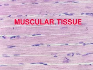
Muscular Tissue
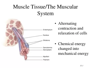
Muscular Tissue. General description 1) components : ---cell: muscle fiber--myofiber elongated thread-liked sarcolemma sarcoplasm SER : sarcoplasmic reticulum ---extracellular G.S: CT with BV, LV and N. 2) classification According to the structure and function

Muscular Tissue
E N D
Presentation Transcript
General description 1) components: ---cell: muscle fiber--myofiber • elongated thread-liked • sarcolemma • sarcoplasm • SER : sarcoplasmic reticulum ---extracellular G.S: CT with BV, LV and N
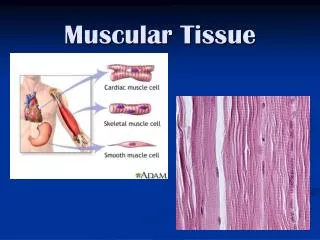
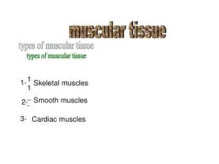
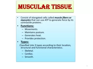
2) classification According to the structure and function • skeletal muscle: striated voluntary m. • cardiac muscle: striated involuntary m. • smooth muscle: unstriated involuntary m. smooth muscle skeletal muscle cardiac muscle
Often called Striated or Voluntary Muscle • Has striations that are visible under light microscopy • Under voluntary control • Attached to skeleton ( skeletal)
Characteristics of Skeletal Muscles Muscle cells are elongated fibers with many peripheral nuclei Each muscle fiber contains many myofibrils Each myofibril contains many microfilaments
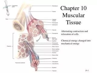
Connective tissue coverings (responsible for force transduction): Epimysium: surround collection of fascicles Perimysium: surround fascicle Endomysium: surround individual fibers
epimysium Perimysium Entire muscle Endomysium Fasciculus of muscle fibres A muscle fibre
1)microstructure of skeletal muscle fiber ①long cylindrical 10-100um in diameter,1-40mm long ②multinucleate, nuclei are ovoid, distributed under sarcolemma ③ filled with longitudinal parallel-arranged myofibrils
Longitudinal section Transverse section
muscle fiber myofibril Thin Filaments Thick Filaments
myofibril: • 1-2um in diameter • cross striation: light band -I band, dark band- A band
--A band: M-line, H-band --I band: Z-line I band A band H-band Z-line M-line
--A band (except H band): Thin Filaments Thick Filaments H band: only Thick Filaments --I band: only Thin Filaments
sacromere sacromere:1/2 I band, A band ,1/2 I band the smallest structural and functional unit of myofibril
2)Ultrastructure of myofiber myofibril: ---thick myofilament: • composed of myosin: ---thin myofilament: • Compose of • A. actin • B. tropomyosin • C.troponin:
Transverse tubule (T tubule): ---definition: sarcolemma and basement membrane invaginate into sarcoplasm to form a transverse distributed tubular system ---location:A-I junctional part ---function: transfer the information into cytoplasm
③Sarcoplasmic reticulum: ---definition: A longitudinal distributed tubular system formed by smooth endoplasmic reticulum Sarcoplasmic reticulum
---structure: • H-A band: longitudinal enclose myofibril-- longitudinal tubule • A-I junction: enlarge to form flattened sac —terminal cisternae • triad: one T-tubule + two terminal cisternae • ---function: there are calcium pump proteins (ATPase) on membrane, so it can store and release calcium ions triad
1) Microstructure of cardiac muscle • ①short column in shaped, 100um long,15um in diameter, with branches, the branches associated with each other to form a network • ②1-2 ovoid nuclei, centrally-located • ③striated, but no very clear • ④intercalated disc: junctional part • LM: dark striation across the cardiac fibers
2) Ultrastructure of cardiac muscle similar to skeletal muscle, composed of thick, thin filament and have sarcomere ①myofibril have different diameter, the boundary of myofibril is not very clear
diad ②transverse tubule are thicker, located at Z-line level (skeletal muscle : A-I junctional part) ③sarcoplasmic reticulum is not well-developed, form less terminal cisternae ④diad: one T-tubule + one terminal cisternae (skeletal muscle : triad) cardiac muscle triad skeletal muscle
transverse portion: connection -intermediate junction -desmosome longitudinal portion: communication -gap junction transverse portion longitudinal portion
microstructure of smooth muscle ①elongated, spindle-shaped cells, 8um in diameter, 200( 20-500)um long ②rob-liked or ovoid nucleus ③no striation, no myofibril
2) ultrastructure of smooth muscle ①caveola on sarcolemme invaginate into cytoplasm ②dense patch: under sarcolemma dense body: in sarcoplasm ③intermediate filament: caveola dense body dense pach
1. The location of T tubules of cardiac muscle differ from those of skeletal muscle, the former lie at the • A. A-I junctions B.I bands • C. A bands D.M lines • E. Z lines √
2. Sarcoplasmic reticulum of muscle fiber is composed of • A. lysosome B. mitochondria • C. microsome D. RER • E. SER √
3.The cell junction which transmit rapidly an electrical impulse between adjacent smooth muscle cells is • A. tight junction B. intermediate junction • C. gap junction D. desmosome • E. semidesmosome √
Questions: • Describe the characteristics of muscle tissue. • Compare the similar with the difference for three kinds of muscle cells according to their fine structure and ultrastructure.