Download
1 / 47
470 likes | 496 Vues
Explore the muscle tissue and the muscular system, including types of muscle tissue, functions, properties, connective tissues, trauma-induced muscle damage, and crush syndrome. Learn about muscle tone, hypertrophy, exercise-induced muscle damage, and more.

E N D
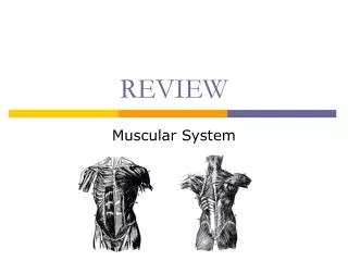
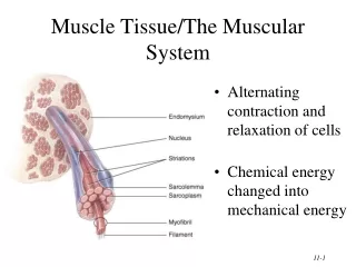
Muscle Tissue/The Muscular System • Alternating contraction and relaxation of cells • Chemical energy changed into mechanical energy
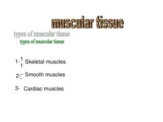
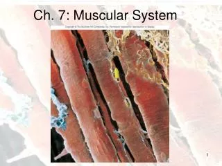
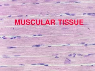
3 Types of Muscle Tissue • Skeletal muscle • attaches to bone, skin or fascia • striated with light & dark bands visible with scope • voluntary control of contraction & relaxation
3 Types of Muscle Tissue • Cardiac muscle • striated in appearance • involuntary control • autorhythmic because of built in pacemaker
3 Types of Muscle Tissue • Smooth muscle • attached to hair follicles in skin • in walls of hollow organs -- blood vessels & GI • nonstriated in appearance • involuntary
Functions of Muscle Tissue • Producing body movements • Stabilizing body positions • Regulating organ volumes • bands of smooth muscle called sphincters • Movement of substances within the body • blood, lymph, urine, air, food and fluids, sperm • Producing heat • involuntary contractions of skeletal muscle (shivering)
Properties of Muscle Tissue • Excitability • respond to chemicals released from nerve cells • Conductivity • ability to propagate electrical signals over membrane • Contractility • ability to shorten and generate force • Extensibility • ability to be stretched without damaging the tissue • Elasticity • ability to return to original shape after being stretched
Skeletal Muscle -- Connective Tissue • Superficial fascia is loose connective tissue & fat underlying the skin • Deep fascia = dense irregular connective tissue around muscle • Connective tissue components of the muscle include • epimysium = surrounds the whole muscle • perimysium = surrounds bundles (fascicles) of 10-100 muscle cells • endomysium = separates individual muscle cells • All these connective tissue layers extend beyond the muscle belly to form the tendon
Atrophy and Hypertrophy • Atrophy • wasting away of muscles • caused by disuse (disuse atrophy) or severing of the nerve supply (denervation atrophy) • the transition to connective tissue can not be reversed • Hypertrophy • increase in the diameter of muscle fibers • resulting from very forceful, repetitive muscular activity and an increase in myofibrils, SR & mitochondria
Exercise-Induced Muscle Damage • Intense exercise can cause muscle damage • electron micrographs reveal torn sarcolemmas, damaged myofibrils an disrupted Z discs • increased blood levels of myoglobin & creatine phosphate found only inside muscle cells • Delayed onset muscle soreness • 12 to 48 Hours after strenuous exercise • stiffness, tenderness and swelling due to microscopic cell damage
Muscle Tone • Involuntary contraction of a small number of motor units (alternately active and inactive in a constantly shifting pattern) • keeps muscles firm even though relaxed • does not produce movement • Essential for maintaining posture (head upright) • Important in maintaining blood pressure • tone of smooth muscles in walls of blood vessels
Trauma Induced Muscle Damage Crush Syndrome-
Case Details • Rural area- • 18 year old patient – 80 Kg • Working on road, rolled 12 ton steamroller into ditch and patient trapped under steamroller • ALS arrival within 15 min- 3 large bore IV started
Case Details Con’t • Patient GCS 15 upon ALS arrival –LOAx3 • BP >160 systolic • HR 90s, colour good, cap refil N • Resp 18 chest clear • Major injuries - ?crushed pelvis ;pelvis and lower extremities not accessible • PMEDHX- unremarkable, no meds
Details: • Crane on site ready to extricate • What are some of the preparations you will do? • What do you anticipate? • What equipment do you need?
Case continuation • Patient extricated and a large 8” piece of metal came out of R femoral area with moving the roller off his pelvis • Immediate arterial bleeding resulted • Direct pressure added to wound by FP • Clinical condition change: -HR increased to 160 from 100 during extrication Colour became poor (pale-cyanotic) BP unpalpable
Treatment Rendered • Direct pressure to wound all the way to trauma center • How to stabilize an unstable pelvis: • KED? • Sager? • Broad bandage to pelvis - ++ tight? • How is it done?
Patient Outcome en route and in Hospital • After 3-4L fluid- HR stabilized at 120 • BP 98/62 • Colour returned somewhat • Arterial bleed continued to be an issue • Direct pressure • In hospital developed rhadbodmyolysis due to crush syndrome
Crush Syndrome and Rhabdomyolysis • "Severe systemic manifestation of trauma and ischemia involving soft tissues, principally skeletal muscle, due to prolonged severe crushing. It leads to increased permeability of the cell membrane and to the release of potassium, enzymes, and myoglobin from within cells and into plasma (rhabdomyolysis). Ischemic renal dysfunction secondary to hypotension and diminished renal perfusion results in acute tubular necrosis and uremia”
Two Types of Smooth Muscle • Visceral (single-unit) • in the walls of hollow viscera & small BV • autorhythmic • gap junctions cause fibers to contract in unison • Multiunit • individual fibers with own motor neuron ending • found in large arteries, large airways, arrector pili muscles,iris & ciliary body
Regeneration of Muscle • Skeletal muscle fibers cannot divide after 1st year • growth is enlargement of existing cells • repair • satellite cells & bone marrow produce some new cells • if not enough numbers---fibrosis occurs most often • Cardiac muscle fibers cannot divide or regenerate • all healing is done by fibrosis (scar formation) • Smooth muscle fibers (regeneration is possible) • cells can grow in size (hypertrophy) • some cells (uterus) can divide (hyperplasia) • new fibers can form from stem cells in BV walls
Aging and Muscle Tissue • Skeletal muscle starts to be replaced by fat beginning at 30 • “use it or lose it” • Slowing of reflexes & decrease in maximal strength • Change in fiber type to slow oxidative fibers may be due to lack of use or may be result of aging
Abnormal Contractions • Spasm = involuntary contraction of single muscle • Cramp = a painful spasm • Tic = involuntary twitching of muscles normally under voluntary control--eyelid or facial muscles • Tremor = rhythmic, involuntary contraction of opposing muscle groups • Fasciculation = involuntary, brief twitch of a motor unit visible under the skin
Muscle Attachment Sites:Origin and Insertion • Skeletal muscles shorten & pull on the bones they are attached to • Origin is the bone that does not move when muscle shortens (normally proximal) • Insertion is the movable bone (some 2 joint muscles) • Fleshy portion of the muscle in between attachment sites = belly
Lever Systems and Leverage • Muscle acts on rigid rod (bone)that moves around a fixed point called a fulcrum • Resistance is weight of bodypart & perhaps an object • Effort or load is work doneby muscle contraction • Mechanical advantage • the muscle whose attachment is farther from the joint will produce the most force • the muscle attaching closer to the joint has the greater range of motion and the faster the speed it can produce
Coordination Within Muscle Groups • Most movement is the result of several muscle working at the same time • Most muscles are arranged in opposing pairs at joints • prime mover or agonist contracts to cause the desired action • antagonist stretches and yields to prime mover • synergists contract to stabilize nearby joints • fixators stabilize the origin of the prime mover • scapula held steady so deltoid can raise arm
How Skeletal Muscle are Named • Direction the muscle fibers run • Size, shape, action, number of origins or locations • Examples from Table 11.2 • triceps brachii -- 3 sites of origin • quadratus femoris -- square shape • serratus anterior -- saw-toothed edge
Muscles of Facial Expression • Arise from skull & insert onto skin • Encircle eyes, nose & mouth • Express emotions • Facial Nerve (VII) • Bell’s palsy = facial paralysis due
Muscles of Facial Expression • Orbicularis oculi closes the eye • Levator palpebrae superioris opens the eye • Orbicularis oris puckers the mouth • Buccinator forms the muscular portion of the cheek & assists in whistling, blowing, sucking & chewing
Muscles that Move the Mandible • Masseter, temporalis & pterygoids • Arise from skull & insert on mandible • Cranial nerve V (trigeminal nerve) • Protracts, elevates or retracts mandible • Temporalis & Masseter elevate the mandible (biting) • temporalis retracts
Muscles that Move the Head • Sternocleidomastoid muscle • arises from sternum & clavicle & inserts onto mastoid process of skull • innervated by cranial nerve XI (spinal accessory) • contraction of both flexes the cervical vertebrae & extends head • contraction of one, laterally flexes the neck and rotates face in opposite direction
Muscles of Abdominal Wall • Notice 4 layers of muscle in the abdominal wall
Muscles of Abdominal Wall • 4 pairs of sheetlike muscles • rectus abdominis = vertically oriented • external & internal obliques and transverses abdominis • wrap around body to form anterior body wall • form rectus sheath and linea alba • Inguinal ligament from anterior superior iliac spine to upper surface of body of pubis • Inguinal canal = passageway from pelvis through body wall musculature opening seen as superficial inguinal ring • Inguinal hernia = rupture or separation of abdominal wall allowing protrusion of part of the small intestine (more common in males)
Muscles Used in Breathing • Breathing requires a change in size of the thorax • During inspiration, thoracic cavity increases in size • external intercostal lift the ribs • diaphragm contracts & dome is flattened • During expiration, thoracic cavity decreases in size • internal intercostal mm used in forced expiration • Diaphragm is innervated by phrenic nerve (C3-C5) but intercostals innervated by thoracic spinal nerves (T2-T12) Quadratus lumborum fills in space between 12th rib & iliac crest to create posterior body wall
Stabilizing the Pectoral Girdle • Anterior thoracic muscles • Subclavius extends from 1st rib to clavicle • Pectoralis minor extends from ribs to coracoid process • Serratus anterior extends from ribs to inner surface of scapula • Posterior thoracic muscle • Trapezius extends from skull & vertebrae to clavicle & scapula • Levator scapulae extends from cervical vertebrae to scapula • Rhomboideus extends from thoracic vertebrae to vertebral border of scapula
Axial Muscles that Move the Arm • Pectoralis major & Latissimus dorsi extend from body wall to humerus.
Muscles that Move the Arm • Deltoid arises from acromion & spine of scapula & inserts on arm • abducts, flexes & extends arm • Rotator cuff muscles extend from scapula posterior to shoulder joint to attach to the humerus • supraspinatus & infraspinatus : above & below spine of scapula • subscapularis on inner surface of scapula
Flexors of the Forearm (elbow) • Cross anterior surface of elbow joint & form flexor muscle compartment • Biceps brachii • scapula to radial tuberosity • flexes shoulder and elbow & supinates hand • Brachialis • humerus to ulna • flexion of elbow • Brachioradialis • humerus to radius • flexes elbow
Extensors of the Forearm (elbow) • Cross posterior surface of elbow joint & forms extensor muscle compartment • Triceps brachii • long head arises scapula • medial & lateral heads from humerus • inserts on ulna • extends elbow & shoulder joints • Anconeus • assists triceps brachii in extending the elbow
Cross-Section Through Forearm • If I am looking down onto this section is it from right or left arm?
Muscles that Move the Vertebrae • Quite complex due to overlap • Erector spinae fibers run longitudinally • 3 groupings • spinalis • iliocostalis • longissimus • extend vertebral column • Smaller, deeper muscles • transversospinalis group • semispinalis, multifidis & rotatores • run from transverse process to dorsal spine of vertebrae above & help rotate vertebrae
Muscles Crossing the Hip Joint • Iliopsoas flexes hip joint • arises lumbar vertebrae & ilium • inserts on lesser trochanter • Quadriceps femoris has 4 heads • Rectus femoris crosses hip • 3 heads arise from femur • all act to extend the knee • Adductor muscles • bring legs together • cross hip joint medially • see next picture • Pulled groin muscle • result of quick sprint activity • stretching or tearing of iliopsoas or adductor muscle
Adductor Muscles of the Thigh • Adductor group of muscle extends from pelvis to linea aspera on posterior surface of femur • pectineus • adductor longus • adductor brevis • gracilis • adductor magnus (hip extensor)
Muscles of the Butt & Thigh • Gluteus muscles • maximus, medius & minimus • maximus extends hip • medius & minimus abduct • Deeper muscles laterally rotate femur • Hamstring muscles • semimembranosus (medial) • semitendinosus (medial) • biceps femoris (lateral) • extend hip & flex knee • Pulled hamstring • tear of origin of muscles from ischial tuberosity
Cross-Section through Thigh • 3 compartments of muscle with unique innervation • anterior compartment is quadriceps femoris innervated by femoral nerve • medial compartment is adductors innervated by obturator nerve • posterior compartment is hamstrings innervated by sciatic nerve
Muscles of the Calf (posterior leg) • 3 muscles insert onto calcaneus • gastrocnemius arises femur • flexes knee and ankle • plantaris & soleus arise from leg • flexes ankle • Deeper mm arise from tibia or fibula • cross ankle joint to insert into foot • tibialis posterior • flexor digitorum longus • flexor hallucis longus • flexing ankle joint & toes
Muscles of the Leg and Foot • Anterior compartment of leg • extensors of ankle & toes • tibialis anterior • extensor digitorum longus • extensor hallucis longus • tendons pass under retinaculum • Shinsplits syndrome • pain or soreness on anterior tibia • running on hard surfaces • Lateral compartment of leg • peroneus mm plantarflex the foot • tendons pass posteriorly to axis of ankle joint and into plantar foot