Muscular System
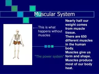
Muscular System. Made up of organs Muscles Remember organs are made up of tissue Primarily skeletal muscle tissue, nervous tissue and blood and other connective tissues And tissue is made up of cells

Muscular System
E N D
Presentation Transcript
MuscularSystem • Made up of organs • Muscles • Remember organs are made up of tissue • Primarily skeletal muscle tissue, nervous tissue and blood and other connective tissues • And tissue is made up of cells • composed of specialized cells that use the chemical energy stored in nutrients to exert a pulling force on structures to which they are attached.
Functions • Voluntary Movement – walking, talking, breathing, smiling (skeletal) • Provide muscle tone (skeletal) • Maintain posture (skeletal) • Propel body fluids and food (smooth) • Distributes heat (smooth) • Generate heart beat (cardiac) • Maintains normal body temperature • Generates 85% of body heat • Compensates for cold by shivering
1 2 Skeletal Skeletal 4 3 Cardiac Smooth
Smooth Muscle Involuntary Surrounding walls of hollow organs and glands Spindle shaped Not striated Single nucleus Central nucleus Gap Junctions Single and Multi-unit Muscle Cell Characteristics
Muscular System • The muscular system consists of skeletal muscles and their associated connective tissues. • It does not include cardiac muscle and smooth muscle, which are associated with the systems in which they are found, such as the cardiovascular, digestive, urinary, or other organ systems. • Individual skeletal muscle is separated from adjacent muscles by dense connective tissue (facia); projects beyond the ends to form a tendon which intertwine with the periosteum of a bone, attaching the two • Tendinitis – inflamed from injury or repeated stress of athletic activity
Skeletal Voluntary Attached to bones Cylindrical Striated Multi-nucleated Nuclei near membrane Tight Junctions Form Motor Units Cardiac Involuntary Heart Cylindrical/Branched Striated Single Nucleus Central Nucleus Gap Junctions – intercalated discs Figure 8 shaped Pacemaker Muscle Cell Characteristics
Muscle Anatomy • Endomysium - connective tissue that surrounds each muscle fiber (cell). • Perimysium – connect. tissue that encircles a group of muscle fibers, forming a fascicle. • Epimysium (fascia)– connect. tissue that encircles all the fascicles to form a complete muscle. • A tendon is fibrous protein, cordlike extension of the preceding three linings. It extends beyond the muscle tissue to connect the muscle to a bone or to other muscles.
Epimysium Perimysium (Myofiber) Wrapped in Endomysium Fasicle (Actin & Myosin) Perimysium
Muscle Cell Anatomy • Myofiber – muscle cell • Should be “myocyte” • Prefix myo = cell *Cylindrical *Multi nucleated *Striated
Muscle Cell Anatomy • Sarcolemma – specialized cell membrane of muscle cell (actively transports Na+ and K+) • Sarcoplasm – cytoplasm of muscle cells (contains most mitochondria of any cell and oval nuclei)
Muscle Cell Anatomy • Sarcoplasmic Reticulum (SR)– specialized SER for storing and releasing and actively transporting calcium - Ca++ • Transverse Tubules (T-tubules)– special passages for Na+ that pass over SR
Muscle Cell Anatomy • Myofibrils – cylindrical organelles that contain the myofilaments needed for muscle contraction • Myofilaments – protein fibers • Thin filaments – actin • Thick filaments – myosin
Muscle Cell Anatomy • Sarcomere – functional unit of contraction; a section of a myofibril • The organization of actin and myosin filaments that make up a myofibril produce the alternating light and dark striations charachteristic of skeletal muscle (and cardiac muscle) • The striations form a repeating pattern of units called sarcomeres.
Sarcomere Anatomy • Z Line – membrane that marks the end of the sarcomere – actin is attached here • I band – at edges of sarcomere – light band – contains only actin • A Band – Dark part of sarcomere – contain myosin (some parts have overlapping actin)
Sarcomere Anatomy • H zone – very center of A band – a little lighter than rest of A band since only contain myosin – no overlapping actin • M line – thickening in the center of the sarcomere, proteins that hold myosin in place
MUSCLE INNERVATION (stimulation) • Motor Unit – One nerve connected to brain or spinal cord and all of the muscle cells/ fibers that it innervates (stimulates)
All or None Principle • All or None Principle – when you contract a motor unit, every fiber or cell in the motor unit contracts and each contracts to the fullest extent • How can you get different strengths of contraction in the same muscle? • # OF MOTOR UNITS ACTIVATED! • Recruitment – multiple motor units stimulated for increased contraction • Precise movement - less motor units recruited (less cells) Ex. Eye movement • Gross movement - more motor units recuited (more cells) Ex. Quadriceps *Remember skeletal muscles have voluntary control.
Skeletal Muscle Contraction • A contraction is movement within the myofibrils, actin & myosin slide past one another, sarcomere shortens • In order for a muscle cell to contract, it must be stimulated by a neuron. • Neuromuscularjunction - where a motor neuron meets a muscle cell and stimulates it.
Neuromuscular Junction • Sarcolemma – cell membrane • Motor end plate – specialized folded region of sarcolemma with neurotransmitter receptors • Synapse - part where muscle membrane meets the nerve (do not actually touch), space where information passes • Axon – cytoplasmic extension of the nerve cell that meets the muscle
Neuromuscular Junction • Neurotransmitter – chemical that allows neuron to communicate will cell • Acetylcholine(Ach) – neurotransmitter that sets off contraction • Synapticvesicle – contains acetylcholine • Synapticcleft – space in between axon and motor end plate where Ach is dumped
Neuromuscular Junction • T-Tubule – when Ach binds to receptors on motor end plate – opens T-tubule channels and allows Na+ to flow in • Terminal cisternae– specialized region of the SR that serve as a reservoire for Ca++ • Sarcomere – contractile unit that extends from one Z line to the next
Components of Muscle Contraction • Myosin – thick filament that pulls actin in to cause contraction • Cross-bridges – globular part of myosin that projects outward and attaches to actin • Actin – thin filament/has binding sites for myosin
Components of Muscle Contraction • Tropomyosin – a rope like protein that wraps around actin covering the active sites on actin so that myosin can’t bind to the actin
Components of Muscle Contraction • Troponin – small proteins that attach to the tropomyosin– has a Ca++ binding site – when Ca++ binds it changes shape and in turn causes the tropomyosin to swivel off of the active sites on actin
Steps of Contraction • Ach is released from the axon into the synaptic cleft due to stimulation by nerve impulse (the brain) • Reminder: Ach is a neurotransmitter that motor neurons use to control skeletal muscle contraction; synthesized in cytoplasm of motor neuron and stored in synaptic vesicle • Ach binds to receptors on the motor end plate • This opens the T tubules – Na+ flow in through the T-tubules
Steps of Contraction • Na+ flowing in through the T-tubules causes channels in SR to open releasing Ca++ • Ca++ attaches to troponin causing it to change shape • The troponin shape change causes the tropomyosin to swivel off of the actin active sites • ATP Activated myosin heads pop up and grab on to actin and swivel forward (power stroke) dragging the actin inward
Steps of Contraction • Many myosin heads are popping up and grabbing on all at once – they are staggered so that some are always attached • This continues as long as there is Calcium present and ATP to power the process (usually don’t run out of the ATP - exhaustion)
What physically happens to the sarcomere during contraction? I band disappears during contraction and so does the H zone as actin is pulled in over myosin (all light areas because actin and myosin completely overlap)
How to Stop a Normal Contraction • This is not exhaustion (running out of ATP) – just normal stopping • Destroy the Ach • Pump out the Na+ • Pump all of the Ca++ back into the SR (active transport with the help of ATP) • ATP is also needed to disconnect head from actin (just as it was needed to make the attachment)
* Latent : Stimulus to triggers Ach to release * Latent: Sodium pumped in * Latent/Contraction: Calcium pumped in contraction * Relaxation: Calcium pumped out • Relaxation/maintaining: • Sodium pumped out Ach breaks down relaxation
Muscle TwitchA single muscle contraction • Latent period – Ach is released, Na+ rushes in, Ca++ is released, active sites are uncovered on actin, myosin binds to actin • Contraction period – myosin is pulling actin inward • Relaxation period – Ca++ is being sucked up by the SR, tropomyosin is recovering active sites on actin, myosin can no longer bind to actin, Na+ is also pumped out and Ach is destroyed in the synaptic cleft
Energy Usage (Muscle Metabolism) • In order for muscles to contract, ATP must be available in the muscle fiber. ATP is available from: • Glycogen stored in muscle cell. Glycogen is broken down to release glucose ATP is then generated from glucose by CR. • Glucose and fatty acids obtained from the bloodstream. When energy requirements are high, glycogen stored in the liver & fatty acids from fat stored in adipose cells & the liver are released into the bloodstream Glucose and fatty acids are then absorbed from the bloodstream by muscle cells ATP is then generated by CR. • Creatine phosphate. A high-energy molecule stored in muscle cells transfers its high-energy phosphate group to ADP to form ATP able to generate enough ATP to maintain muscle contraction for about 15 seconds.
Aerobic Respiration • Needs a lot of oxygen to burn glucose this way and make ATP • Makes 38 ATP/glucose – only efficient way to make enough ATP for sustained muscle contraction • Myoglobin – red protein in muscle that binds oxygen and stores it • Why need oxygen? • Make ATP aerobically • Reload myoglobin • Replenish creatine phosphate
Aerobic Respiration • Advantages: Aerobic respiration generates a large amount of ATP. • Disadvantages: Aerobic respiration is relatively slow and requires oxygen.
Anerobic Respiration • Oxygen debt – can’t get enough , can’t make enough ATP – feel fatigued, breathe heavily • Glycolysis/Fermentation – only make 2 ATP/glucose and lactic acid • No activity can be totally anaerobic and be compatible with life • Your need for ATP is greater than you can supply the oxygen for • Anaerobic activities include anything that makes you breathe excessively heavy – sign you’re not getting enough oxygen • Sprinting, swimming really fast, biking really fast, lifting really heavy weights
Anaerobic Respiration • Advantages: Anaerobic respiration is relatively rapid, and does not require oxygen. • Disadvantages: Anaerobic respiration generates only two ATPs and produces lactic acid which contributes to muscle fatigue. Can’t keep this process up very long (will run out of ATP). After exercise, the liver and muscles need to convert the lactic acid back to pyruvic acid. In order to do that, a lot of the oxygen the body is now taking in does the conversion instead of being used elsewhere. This is known as “repaying the debt,” hence the term “oxygen debt.”