Download
1 / 43
430 likes | 571 Vues
Discover the composition, organization, and role of skeletal muscle tissue. Learn about muscle fibers, connective tissues, and the sarcomere structure in this comprehensive overview.

E N D
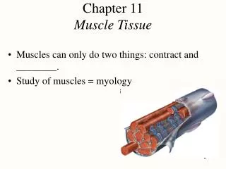
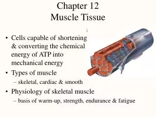
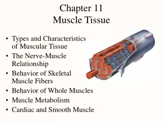
Muscle Tissue: Introduction • Muscle tissue is composed of cells differentiated for optimal use of the universal cell property termed contractility. • Microfilaments and associated proteins together generate the forces necessary for cellular contraction. • Nearly all muscle cells are of mesodermal origin and they differentiate mainly by a gradual process of cell lengthening with simultaneous synthesis of myofibrillar proteins. • Three types of muscle tissue can be distinguished on the basis of morphologic and functional characteristics and the structure of each type is adapted to its physiologic role. • Skeletal muscle. • Cardiac muscle. • Smooth muscle
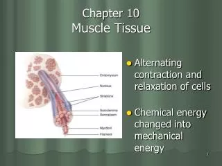
Skeletal Muscle • Skeletal muscle consists of muscle fibers, which are long, cylindrical multinucleated cells with diameters of 10–100 m. • Multinucleation results from the fusion of embryonic mesenchymal cells called myoblasts. • The long oval nuclei are usually found at the periphery of the cell under the cell membrane. • This characteristic nuclear location is helpful in discriminating skeletal muscle from cardiac and smooth muscle, both of which have centrally located nuclei.
Organization • The masses of fibers that make up the various types of muscle are arranged in regular bundles surrounded by the Epimysium: an external sheath of dense connective tissue surrounding the entire muscle. The connective tissue (thin septa of connective tissue from the epimysium) surrounding each fascicle (bundles of fibers within a muscle) is called the Perimysium. • Each muscle fiber is itself surrounded by a more delicate connective tissue, the Endomysium: composed of a basal lamina synthesized by the multinucleated fibers themselves as well as reticular fibers and fibroblasts. • Within each fiber the nuclei are displaced peripherally against the sarcolemma.
The Role of The Connective Tissue • Is to transmit the mechanical forces generated by the contracting muscle cell/fibers because individual muscle cells seldom extend from one end of a muscle to the other. • Blood vessels penetrate the muscle within the connective tissue septa and form a rich capillary network in the endomysium.
Myotendinous junction. - Tendons develop together with skeletal muscles and join muscles to the periosteum of bones. The collagen fibers of tendons are continuous with those in the connective tissue layers in the muscle, forming a strong unit that allows muscle contraction to move the skeleton. - The longitudinal section shows part of a tendon (T) inserted into the endomysium and perimysium of a muscle.
Muscle Fibers • Under the light microscope, longitudinally sectioned skeletal muscle fibers show cross-striations of (alternating light and dark bands. • The darker bands are called A bands. • The lighter bands are called I bands . • In the TEM each I band is seen to be bisected by a dark transverse line, the Z line . • The repetitive functional subunit of the contractile apparatus, the Sarcomere, extends from Z line to Z line.
Muscle Fibers • The sarcoplasm has little RER or free ribosomes. • It is filled primarily with long cylindrical filamentous bundles called myofibrils running parallel to the long axis of the fiber. • The myofibrils have a diameter of 1–2 m and consist of an end-to-end repetitive arrangement of sarcomeres. The lateral registration of sarcomeres in adjacent myofibrils causes the entire muscle fiber to exhibit a characteristic pattern of transverse striations. • The A and I banding pattern in sarcomeres is due mainly to the regular arrangement of two types of myofilaments: Thick&thin.
The Thick & Thin filaments • The Think filaments (1.6 m long and 15 nm wide), they occupy the A band, the central portion of the sarcomere. The thin filaments run between and parallel to the thick filaments and have one end attached to the Z line. • Thin filaments are (1.0 m long and 8 nm wide). • The I bands consist of the portions of the thin filaments that do not overlap the thick filaments (which is why they are lighter staining). • The A bands are composed mainly of thick filaments in addition to overlapping portions of thin filaments. • The A band shows the presence of a lighter zone in its center, the H zone, that corresponds to a region consisting only of the rod-like portions of the myosin molecule with no thin filaments present. • Bisecting the H zone is the M line , a region where lateral connections are made between adjacent thick filaments. Major proteins present in the M line region are myomesin.
The Thick & Thin filaments • Thin filaments are composed of F-actin, the tropomyosin, which also and troponin. • Thick filaments consist primarily of myosin. • Myosin and actin together represent 55% of the total protein of striated muscle.
Sarcoplasmic Reticulum & Transverse Tubule System • In muscle the smooth endoplasmic reticulum (SER) is specialized for Ca2+ ion sequestration. • To provide for a uniform contraction, skeletal muscle fibers have a system of transverse (T) tubules. These fingerlike invaginations of the sarcolemma form a complex network of tubules that encircles every myofibril near the A-I band boundaries of each sarcomere. • Adjacent to opposite sides of each T tubule are expanded terminal cisternae of the sarcoplasmic reticulum. This specialized complex, consisting of a T tubule and usually two small cisternae of sarcoplasmic reticulum, is known as a Triad. • At the triad, depolarization of the sarcolemma-derived T tubules is transmitted to the sarcoplasmic reticulum membrane.
Cardiac Muscle • During embryonic development, the mesoderm cells of the primitive heart tube align into chainlike arrays. • Cardiac muscle cells form complex junctions between extended processes. • Cells within a fiber often branch and bind to cells in adjacent fibers. • The heart consists of tightly knit bundles of cells, interwoven in a fashion that provides for a characteristic wave of contraction that leads to a wringing out of the heart ventricles.
Cardiac Muscle • Mature cardiac muscle cells are approximately 15 m in diameter and from 85 to 100 m in length. • They exhibit a cross-striated banding pattern comparable to that of skeletal muscle. • Each cardiac muscle cell possesses only one or two centrally located pale-staining nuclei. • Surrounding the muscle cells is a delicate sheath of endomysium containing a rich capillary network.
Cardiac Muscle • A unique characteristic of cardiac muscle is the presence of dark-staining transverse lines that cross the chains of cardiac cells at irregular intervals. • These intercalated discs represent the interface between adjacent muscle cells where many junctional complexes are present. • Transverse regions of these steplike discs have many desmosomes and fascia adherentes (which resemble the zonula adherentes between epithelial cells) and together • These junctions serve to bind cardiac cells firmly together to prevent their pulling apart under constant contractile activity. • The more longitudinal portions of each disc have multiple gap junctions, which provide ionic continuity between adjacent cells. • These act as "electrical synapses" and allow cells of cardiac muscle to act as in a multinucleated syncytium, with contraction signals passing in a wave from cell to cell.
Cardiac Muscle • The structure and function of the contractile proteins in cardiac cells are essentially the same as in skeletal muscle. • The T tubule system and sarcoplasmic reticulum are not as regularly arranged in cardiac fiber. • The T tubules are more numerous and larger in cardiac muscle than in skeletal muscle and the sarcoplasmic reticulum is less well developed. • Cardiac muscle cells contain numerous mitochondria, which occupy 40% or more of the cytoplasmic volume, reflecting the need for continuous aerobic metabolism in heart muscle. • By comparison, only about 2% of skeletal muscle fiber is occupied by mitochondria.
Smooth Muscle • Smooth muscle fibers are elongated, tapering, and nonstriated cells, each of which is enclosed by a thin basal lamina and a fine network of reticular fibers. • The connective tissues serve to combine the forces generated by each smooth muscle fiber into a concerted action, eg, peristalsis in the intestine. • Smooth muscle cells may range in length from 20 m in small blood vessels to 500 m in the pregnant uterus. • Each cell has a single nucleus located in the center of the cell's broadest part. • To achieve the tightest packing, the narrow part of one cell lies adjacent to the broad parts of neighboring cells.
Smooth Muscle • Concentrated near the nucleus are mitochondria, polyribosomes, cisternae of rough ER, and the Golgi apparatus. • A rudimentary sarcoplasmic reticulum is present in smooth muscle cells, but T tubules are not. • The characteristic contractile activity of smooth muscle is related to the structure and organization of its actin and myosin filaments, which DO NOT exhibit the organization present in striated muscles. • In smooth muscle cells, bundles of thin and thick myofilaments crisscross obliquely through the cell, forming a latticelike network. • The thin filaments of smooth muscle cells lack troponin complexes and instead utilize calmodulin.
Regeneration of Muscle Tissue • The three types of adult muscle have different potentials for regeneration after injury. • In skeletal muscle, although the nuclei are incapable of undergoing mitosis, the tissue can undergo limited regeneration. The source of regenerating cells is the sparse population of mesenchymal satellite cells that lies within the external lamina of each mature muscle fiber. The regenerative capacity of skeletal muscle is limited, however, after major muscle trauma or degeneration. • Cardiac muscle lacks satellite cells and has virtually no regenerative capacity beyond early childhood. • Smooth muscle, composed of simpler, mononucleated cells, is capable of a more active regenerative response. After injury, viable smooth muscle cells undergo mitosis and replace the damaged tissue.