Download
1 / 98
980 likes | 1.01k Vues
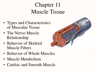
Chapter 11 Muscle Tissue. Types and Characteristics of Muscular Tissue The Nerve-Muscle Relationship Behavior of Skeletal Muscle Fibers Behavior of Whole Muscles Muscle Metabolism Cardiac and Smooth Muscle. Objectives.

E N D
Chapter 11Muscle Tissue • Types and Characteristics of Muscular Tissue • The Nerve-Muscle Relationship • Behavior of Skeletal Muscle Fibers • Behavior of Whole Muscles • Muscle Metabolism • Cardiac and Smooth Muscle
Objectives • Relate the form to the function of the molecular components of muscles. • Identify 3 types of muscle cells from pictures • Explain the mechanisms of contraction • Describe genetic components of muscle growth • Describe aspects of Muscular Dystrophy • Pump you up
Dissections comparing normal, heterozygous and homozygous mice
Myostatin • A growth factor (hormone) that surpresses muscle growth. • Slows down development of muscle stem cells • In 2002, researchers at the University of Pennsylvania showed that monoclonal antibody specific to myostatin improves the condition of mice with muscular dystrophy, presumably by blocking myostatin's action.
Muscular Dystrophy • Group of hereditary diseases in which skeletal muscles degenerate & are replaced with adipose • Etiology: Mainly a disease of males • What types of genetic diseases are primarily found in males? • Sex linked, This is a gene on the X –Chromosome. • appears as child begins to walk • Normal allele makes dystrophin, a protein that links actin filaments to cell membrane • absence of dystrophin leads to torn cell membranes
Muscular Dystrophy Pedigree XM normal Xm MD Y- no allele What is the genotype of Individauals A B C D Who is a carrier?
Duchenne's Muscular Dystrophy • Prevalence estimated pop. of people who are managing it at any given time. • USA: 43,000 • Incedence: the annual diagnosis rate/ number of new cases diagnosed each year. • About 1 in 3,000 • Prognosis: No know cures, only treatments to alleviate suffering and control the rate.
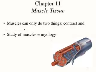
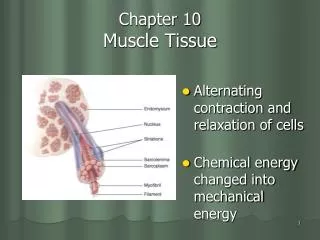
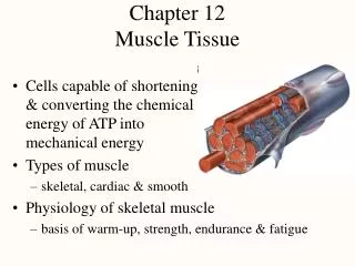
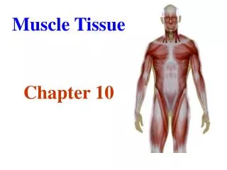
Introduction to Muscle • Movement is a fundamental characteristics of all living things • Cells capable of shortening & transducing the chemical energy of ATP into mechanical energy • Types of muscle • skeletal • cardiac • smooth • Physiology of skeletal muscle • basis of warm-up, strength, endurance & fatigue
Smooth Muscle • Cells spindle shaped • Uninucleated
Cardiac Cells • Shorter and thicker than skeletal • Can keep their own beat, because of nearby pacemaker cells
Universal Characteristics of Muscle • Responsiveness (excitability) • capable of response to chemical signals, stretch or other signals & responding with electrical changes across the plasma membrane • Conductivity • local electrical change triggers a wave of excitation that travels along the muscle fiber • Contractility -- shortens when stimulated • Extensibility -- capable of being stretched • Elasticity -- returns to its original resting length after being stretched
Skeletal Muscle • Voluntary striated muscle attached to bones • Muscle fibers (myofibers) as long as 30 cm • Exhibits alternating light and dark transverse bands or striations • reflects overlapping arrangement of internal contractile proteins • Under conscious control
Connective Tissue Elements of Muscle • Found between muscle fiber and bone or other attachment • endomysium, perimysium, epimysium, fascia, tendon • Not excitable or contractile, but are somewhat extensible & elastic • stretches slightly under tension and recoils when released • Called series-elastic components • are connected to each other in linear series • help return muscles to their resting lengths • adds significantly to power output and efficiency of muscles
Why Multinucleation? • Physically: You need a long tube • Problems of regulation: Takes fewer neurons to synchronize fewer, larger cells • Multinucleation overcomes problems of diffusion because incoming chemicals can always find a control center.
Muscle Fibers (Form follows Function) • Multiple flattened nuclei against inside of plasma membrane • due to fusion of multiple myoblasts during development • unfused satellite cells nearby can multiply to produce a small number of new myofibers • Sarcolemma has tunnel-like infoldings or transverse (T) tubules that penetrate the cell • carry electric current to cell interior • Sarcoplasm is filled with • myofibrils (bundles of parallel protein microfilaments called myofilaments) • glycogen for stored energy & myoglobin binding oxygen • Sarcoplasmic reticulum is series of interconnected, dilated, calcium storage sacs called terminal cisternae
Thick Filaments • Made of 200 to 500 myosin molecules • 2 entwined polypeptides (golf clubs) • Arranged in a bundle with heads (cross bridges) directed outward in a spiral array around the bundled tails • central area is a bare zone with no heads
Thin Filaments • Two intertwined strands of fibrous (F) actin • each subunit is a globular (G) actin with an active site • Groove holds tropomyosin molecules, each blocking the active sites of 6 or 7 G actins • One small, calcium-binding troponin molecule stuck to each tropomyosin molecule
Elastic Filaments • Huge springy protein called titin (connectin) • runs through core of each thick filament • connects thick filament to Z disc structure • Functions • keep thick & thin filaments aligned with each other • resist overstretching • help the cell recoil to its resting length (elasticity)
Regulatory & Contractile Proteins • Myosin & actin are contractile proteins (they do work) • Tropomyosin & troponin are regulatory proteins • act like a switch that starts & stops shortening of muscle cell • the release of calcium into sarcoplasm and its binding to troponin, activates contraction • troponin moves the tropomyosin off the actin active sites
I A I Striations = Organization of Filaments • Dark A bands (regions) alternating with lighter I bands (regions) • anisotrophic (A) and isotropic (I) stand for the way these regions affect polarized light • A band is thick filament region • lighter, central H band area contains no thin filaments • I band is thin filament region • bisected by Z disc protein called connectin, anchoring elastic & thin filaments • from one Z disc (Z line) to the next is a sarcomere
Relaxed versus Contracted Sarcomere • Muscle cells shorten because their individual sarcomeres shorten • pulling Z discs closer together • pulls on sarcolemma • Notice neither thick nor thick filaments change length during shortening • Their overlap changes as sarcomeres shorten
Skeletal muscles contract according to the sliding-filament model: An action potential reaches the axon of the motor neuron. The action potential activates voltage gated calcium ion channels on the axon, and calcium rushes in. The calcium causes acetylcholine vesicles in the axon to fuse with the membrane, releasing the acetylcholine into the cleft between the axon and the motor end plate of the muscle fiber. The acetylcholine diffuses across the cleft and binds to nicotinic receptors on the motor end plate, opening channels in the membrane for sodium and potassium. Sodium rushes in, and potassium rushes out. However, because sodium is more permeable, the muscle fiber membrane becomes more positively charged, triggering an action potential. The action potential spreads through the muscle fiber's network of T tubules, depolarizing the inner portion of the muscle fiber. The depolarization activates voltage-gated calcium channels in the T tubule membrane, which are in close proximity to calcium-release channels in the adjacent sarcoplasmic reticulum. Activated voltage-gated calcium channels physically interact with calcium-release channels to activate them, causing the sarcoplasmic reticulum to release calcium. The calcium binds to the troponin C present on the thin filaments of the myofibrils. The troponin then allosterically modulates the tropomyosin. Normally the tropomyosin sterically obstructs binding sites for myosin on the thin filament; once calcium binds to the troponin C and causes an allosteric change in the troponin protein troponin T allows tropomyosin to move, unblocking the binding sites. Myosin (which is bound to ADP and is in a ready state) binds to the newly uncovered binding sites on the thin filament. It then releases ADP and an inorganic phosphate and delivers a power stroke. Myosin is now bound to actin in the strong binding state. ATP binds myosin, allowing it to release actin and be in the weak binding state. (A lack of ATP makes this step impossible, resulting in rigor mortis.) The myosin then hydrolyzes the ATP and uses the enery to move into the "cocked back" state while releasing ADP and inorganic phosphate. Steps 7 and 8 repeat as long as ATP is available and calcium is present on thin filament. All the while, the calcium is actively pumped back into the sarcoplasmic reticulum. When calcium is no longer present on the thin filament, the tropomyosin changes conformation back to its previous state so as to block the binding sites again. The myosin ceases binding to the thin filament, and the contractions cease. The calcium ions leave the troponin molecule in order to maintain the calcium ion concentration in the sarcoplasm. The active pumping of calcium ions into the sarcoplasmic reticulum creates a deficiency in the fluid around the myofibrils. This causes the removal of calcium ions from the troponin. Thus the tropomyosin-troponin complex again covers the binding sites on the actin fiaments and contraction ceases.
Why warm up? • Release of Adrenalin makes blood vessels dialate, delivering more oxygen. • Increased temperature benefits • More viscous blood • Faster enzyme action • Hemoglobin delivers oxygen faster • Psychological benefit • Example New Zealand Rugby team, The All Blacks perform a Maori war dance (Haka) as part of their warm up
What do you mean by stronger? • 2 TYPES of MUSCLE FIBERS • determined both genetically & functionally • based upon how fast they can produce a contractile twitch • every muscle composed of varying % composition of two types • Type I, slow twitch, Dark meat, endurance • Type II, fast twitch, Light meat, speed
Mark Allen: first male to win 5 consecutive Iron Man Triathlons Held in Hawaii Excellent example of high percentage of slow twitch Type I dark meat Like birds or sea going mammals have.
Carl Lewis • Ran 100 m in 9.86s • 1991 world record • Jumps over cars • 9 Gold Medals • Excellent example of Type II fast twitch muscle • Also a vegan • Outspoken, even damning of those who used performance enhancers. • Like Ben Johnson
Ben Johnson • Between 1968 and 1983 the 100m record was shaved by 0.04s • In one year Johnson beat it by 0.16s • 1988 Seoul Olympics • Johnson: 9.76 s • 27 mph • 45 strides • Ahead the whole time • Lewis sets an American record with 9.92s, but still second • Busted for doping
Johnson’s medal revoked and given to Lewis 5 years later in 2003 Lewis admits he was busted 3 times in 1988 for banned stimulants. He “thought they were herbal supplements” The U.S. Olympic committee found his ingestion to be, “inadvertant” Since it was after the 3 year statute of limitations he could keep his medals Stimulants could have masked more serious steroids from tests Johnson beating Lewis in ‘88 Back to Carl Lewis
Which leads to the question how do steroids work? • Androgenic: increasing masculine traits • Anabolic: building muscles as opposed to • Catabolic: breaking down nutrients
Testosterone • Testosterone: best known natural steroid • Gets to cell, gets into cell, changed to DHT (Dihydrotestsoterone), DHT goes into Nucleus, binds to DNA and starts transcriptional activities to build more muscle mass. • Side effects include decreased sexual function, baldness, acne and Gynecomastia
Nerve-Muscle Relationships • Skeletal muscle must be stimulated by a nerve or it will not contract (paralyzed) • Cell bodies of somatic motor neurons are in brainstem or spinal cord • Axons of somatic motor neurons are called somatic motor fibers • each branches, on average, into 200 terminal branches that supply one muscle fiber each • Each motor neuron and all the muscle fibers it innervates are called a motor unit
Motor Units • A motor neuron & the muscle fibers it innervates • dispersed throughout the muscle • when contract together causes weak contraction over wide area • provides ability to sustain long-term contraction as motor units take turns resting (postural control) • Fine control • small motor units contain as few as 20 muscle fibers per nerve fiber • eye muscles • Strength control • gastrocnemius muscle has 1000 fibers per nerve fiber
Neuromuscular Junctions (Synapse) • region where a nerve fiber makes a functional connection with its target cell (NMJ) • Neurotransmitter (acetylcholine/ACh) released from nerve fiber causes stimulation of muscle cell • Components of synapse • synaptic knob is swollen end of nerve fiber (contains ACh) • motor end plate is specialized region of muscle cell surface • has ACh receptors on junctional folds which bind ACh released from nerve • acetylcholinesterase is enzyme that breaks down ACh & causes relaxation • synaptic cleft = tiny gap between nerve and muscle cells • schwann cell envelopes & isolates NMJ