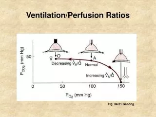
Perfusion
This guide provides an in-depth exploration of the heart's anatomy, layers, chambers, and valves, as well as essential cardiac functions and sounds. Learn about the normal functioning of the heart, including the cardiac cycle, blood circulation (pulmonary and systemic), and the conduction system. We delve into the significance of heart sounds, the impact of developmental changes, and common cardiovascular issues like congenital defects and aging. This resource is essential for understanding cardiac output, assessments, and relevant diagnostic tests for effective healthcare management.

Perfusion
E N D
Presentation Transcript
22 Perfusion
Normal Presentation Heart Pericardium Layers of the heart wall Epicardium Myocardium Endocardium
Figure 22-3 The internal anatomy of the heart, frontal section.
Chambers and Valves of the Heart 4 hollow chambers Right atrium Left atruim Right ventricle Left ventricle Heart chambers separated by valves Atria separated by atrioventricular valves Pulmonic valve Aortic valve
Normal Heart Sounds Closure of valves creates heart sounds AV valve produces S1 Semilunar produces S2 Contraction and relaxation of the heart Splitting of S2 normal in some S3 ventricular gallop S4 atrial gallop Characteristics of heart sounds
Normal Heart Sounds, continued Additional Heart Sounds Damaged tissue Mitral stenosis Ejection clicks Nonejection clicks Friction rubs Heart murmurs
Systemic, Pulmonary, Coronary Circulation Pulmonary circulation Right side of heart Systemic circulation Left side of heart Coronary circulation Vessels that supply the heart muscle itself
Transition from Fetal to Pulmonary Circulation Flow of blood Foramen ovale Blood pumped into aorta, systemic circulation System vascular resistance
The Cardiac Cycle and Cardiac Output One heartbeat of contraction and relaxation Stroke volume Cardiac output Ejection fraction
The Cardiac Cycle and Cardiac Output, continued Cardiac output determined by Autonomic stimulation Preload Afterload Contractility Clinical indicators of cardiac output Cardiac index
Conduction System of the Heart Sinoatrial node (SA node) Creates impulse Depolarization causes myocardial contraction Action potential is basis for EKG Depolarization Repolarization Anatomic landmarks for cardiovascular assessment
Pulse Wave of blood created by LV contraction Peripheral pulse Apical pulse Factors affecting pulse
Blood Pressure Measure of pressure of blood flow through arteries Systolic Diastolic Pulse pressure Measured in millimeters of mercury Determinant of blood pressure Factors affecting blood pressure
Developmental Aspects Pediatric differences Cardiac functioning Oxygenation Cardiovascular changes in pregnancy Cardiac output increases early in pregnancy Enlarging uterus places pressure on veins Interferes with returning blood flow When supine, pressure on vena cava
Figure 22-13 Vena caval syndrome. The gravid uterus compresses the vena cava when the woman is supine. Thisreduces the blood flow returning to the heart and may cause maternal hypotension.
Developmental Aspects Cardiovascular changes in pregnancy, continued Blood volume increases Leukocyte production increases Fibrin and plasma fibrinogen increase
Developmental Aspects, continued Normal changes of aging Determined by genetics Physical, social environment Cardiovascular system Vascular system Pulmonary system Renal system
Figure 22-14 Normal changes of aging in the cardiovascular system.
Alterations Alterations in pediatric cardiology Congenital heart defects Congenital anomalies Common cardiovascular illnesses of aging CAD – Cardiomyopathy Dysrhythmia – Valve disease Shock – Hypertension Stroke – PIH
Assessment Physical assessment Inspection Palpation Percussion Auscultation Perfusion assessments Apical impulse – Heart sounds Cardiac rate and rhythm – Murmurs
Assessing the Pulse Techniques Assess for Rate – Rhythm Volume – Arterial wall elasticity Developmental considerations Pulse sites Apical pulse assessment Apical-radial pulse assessment
Assessing Blood Pressure Measured with sphygmomanometer Blood pressure sites Methods Direct Indirect Auscultate, palpate Korotkoff’s sounds 5 phases of blood pressure
Assessing Blood Pressure, continued Developmental considerations Care settings Common errors in assessing blood pressure Haste Unconscious bias Hypotension Orthostatic hypotension
Health Assessment Interview Explore chief complaint Assessing chest pain Genetic considerations
Diagnostic Tests Determining cardiac function Serum cholesterol, triglycerides, lipids Stress/exercise tests X-ray, MRI, CT, PET Echocardiogram Transesophageal echocardiogram Cardiac catheterization, angiography Pericardiocentesis
Diagnostic Tests, continued Electrocardiography Interpreting an ECG Troponin, MB isoenzyme of creatine kinase Nursing responsibilities Explain the procedure and preparation Assess for medication use Support client during test if necessary Document procedure, monitor results
Caring Interventions Support, improve, promote perfusion Interventions to Reduce stress on the heart Decrease cardiac workload Increase efficacy of cardiac contractions Meet fluid needs Primary prevention Diet Exercise
Caring Interventions, continued Interventions – 3 categories Inputs Outputs Pressure supports Specific skills Assessing pulses Recording and interpreting an ECG Administering CPR
Caring Interventions Specific skills, continued Achieving defibrillation Measuring for, application of elastic compression stockings Fetal monitoring Applying SCD Applying continuous cardio-respiratory monitoring Obtaining capillary wedge pressures
Caring Interventions Specific skills, continued Measuring cardiac output Reading central and peripheral blood pressures Caring for arterial pressure monitoring
Pharmacologic Therapies Classifications Statins Antihypertensives Adrenergic agonists Calcium channel blockers Cardiac glycosides Phosphodiesterase inhibitors Nitrates Thrombolytics