Download
1 / 24
240 likes | 259 Vues
This case study presents a 3-year-old boy with exopthalmos, a mesenchymal lesion invading the orbital space. The diagnosis, differential diagnosis, and staining techniques used for Ewings Sarcoma are discussed.

E N D
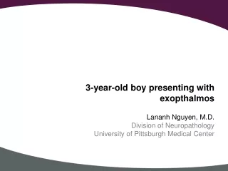
3-year-old boy presenting with exopthalmos Lananh Nguyen, M.D. Division of Neuropathology University of Pittsburgh Medical Center
Clinical history • Patient presented to the PCP complaining of right eye swelling. Given the patient’s history of allergies, antihistamines were prescribed without improvement. • A few weeks afterwards, patient presented to the ED with right eye protrusion and erythema. • Physical exam showed intact vision. • And imaging was performed.
Radiology: Identify the lesion and name the 3 imaging modalities used below.
Radiology: Imaging of the skull lesion. Identify the lesion and name the 3 imaging modalities used below. T1 T1 with contrast T2 This is an extraaxial (nonbrain) mesenchymal lesion invading into the orbital space
A biopsy was performed and an intraoperative consultation was requested. Click on the link for the whole slide image of the smear , scan the virtual slide and try to formulated a differential diagnosis.
A biopsy was performed and an intraoperative consultation was requested. What do you see on the smear? Low power
A biopsy was performed and an intraoperative consultation was requested. What do you see on the smear? Low power It is lesional and abnormally hypercellular
High power smear. What do you see on the smear? Benign or malignant? High power
High power smear. What do you see on the smear? Benign or malignant? High power Small blue cells Mitosis
These are permanent H&E slides. High power
Is it benign or malignant? • Malignant
What is your differential diagnosis? • Small round blue cell tumor • Ewings Sarcoma/ Primitive neuroectodermal tumor • Neuroblastoma • Rhabdomyosarcoma • CNS Primitive neuroectodermal tumor • Lymphoma • Atypical teratoid rhabdoid tumor • Ependymoma
These are the stains the pathologist ordered. • Ewings Sarcoma/ Primitive neuroectodermal tumor – CD99 • Neuroblastoma – Pgp 9.5, synaptophysin or chromogranin • Rhabdomyosarcoma – myogenin, desmin, smooth muscle actin, vimentin • CNS Primitive neuroectodermal tumor – GFAP • Lymphoma – CD3, CD20 • Atypical teratoid rhabdoid tumor – INI • Ependymoma – EMA, p53
Immunohistochemical stains synaptophysin
What do you see on the stains? • CD99 was strongly and diffusely positive • Synaptophysin showed diffuse but patchy cytoplasmic staining • Vimentin (not shown) highlighted vessels • INI (not shown) was intact • All other stains (not shown) in panel were negative
What is your final diagnosis? • Final diagnosis: • Ewings Sarcoma/Primitive Neuroectodermal tumor • FISH studies were positive for t(11;22).
Discussion • Many translocations have been identified for Ewing’s sarcoma. The table lists the most commonly identified ones with t(11;22) as the most common. Adapted from
Discussion • The prognostic factors for increased survival and response to treatment are: • Female gender • Younger children (<10 years old) • Small tumor size or volume • Tumor location (axial worse than extremities) • Decreased serum lactate dehydrogenase (LDH) • No metastasis • Lack of overexpression of p53 • Low Ki67 proliferation index • The 5-year survival rate has increased over the same time from 59% to 76% for children younger than 15 years and from 20% to 49% for adolescents aged 15 to 19 years.[Smith MA, Seibel NL, Altekruse SF, et al.: Outcomes for children and adolescents with cancer: challenges for the twenty-first century. J Clin Oncol 28 (15): 2625-34, 2010.]