Download

1 / 44
460 likes | 744 Vues
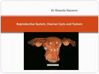
Reproductive System, Ovarian Cysts and Tumors Emad Raddaoui , FCAP, FASC. Ovaries. Ovarian mass Can be either : - Physiological or Neoplastic - Benign or Malignant The most important medical problems in ovaries are the neoplasms

E N D
Reproductive System, Ovarian Cysts and TumorsEmadRaddaoui, FCAP, FASC
Ovaries Ovarian mass Can be either : - Physiological or Neoplastic - Benign orMalignant • The most important medical problems in ovaries are the neoplasms • Death from ovarian cancers is more than that of cervix and uterus together • Silent growth of ovarian tumors is the rule ,which make them so dangerous
Ovarian Cysts and Tumors • Non neoplastic cysts are common but they are not serious problems • Primary inflammation of ovaries is rare • Salpingitis of fallopian tubes frequently causes periovarian reaction (Salpingo-Oophoritis)
Non-Neoplastic and Functional Cysts of ovary • Non Neoplastic Cyst are more common than the neoplastic . • Corpus Luteum and Follicular cystsphysiological cysts. • A corpus luteum cyst results from delayed resolution of a corpus luteum’s central cavity and hemorrhage into this persistent mature corpus luteum. • This condition is self-limited.
Are thin walled fluid filled structures lined internallyby granulosa cells and externally by theca internacells.They arise from the ovarian follicles and are less than 5 cmAre due to distension of un-ruptured graafian follicle. Follicular cysts: Occasionally such cysts may reach several centimeters in size and, if they rupture, can cause abdominal pain.
Chocolate/ Endometriotic cyst • Chocolate cyst is a blood containing cyst resulting from endometriosis with hemorrhage.
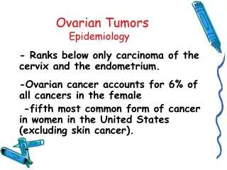
Ovarian Neoplasms- Introduction • The fifth most common cancer in US women • 80% are benign – young (20-45) • 20% are Malignant - older (>40) • 6% of all cancers in women. • 50% deaths due to late detection.
Ovarian Neoplasms: Risk Factors • Null parity • Gonadal Dysgenesis • Family History • Ovarian cancer genes • BRCA1 (17q12) & BRCA2(13q12) (Cancer suppressor, Breast & ovary)
Ovarian Tumors Because the three major cell types make up the normal ovary, there are >25 types of ovarian tumors each with many variants
Ovarian Tumors classification There are THREE main types of primary ovarian tumors: • Epithelial Surface Tm : arederived from the cells on the surface of the ovary. This is the most common form of ovarian cancer and occurs primarily in adults. • Germ Cell Tm :ovarian tumors are derived from the egg producing cells within the body of the ovary. This occurs primarily in children and teens and is rare by comparison to epithelial ovarian tumors. • Sex Cord Tm :ovarian tumors are also rare in comparison to epithelial tumors and this class of tumors often produces steroid hormones. In addition cancers derived from other organs can also spread to the ovaries Secondary /Metastatic Tumors
Surface Epithelial tumors: • Most common primary neoplasms • 90% of malignant tumors of ovary • Serous (tubal) • Mucinous (endocervical& intestinal) • Endometrioid • Transitional cell - Brenners. • Morphologically • Cystic – eg:Cystadenomas • Solid/cystic –eg: Cystadenofibromas • Solid – eg:adenofibromas
Surface Epithelial tumors All types can be benign, borderline , or malignant • Benign : -gross: mostly cystic microscopic:fine papillae, single layer covering (no stratification), no nuclear atypia, no stromal invasion • Borderline :gross: cystic / solid foci microscopic; papillary complexity, stratification, nuclear atypia, no stromal invasion • Malignant: gross: mostly solid & hemorrhage / necrosis microscopic:papillary complexity, stratification, nuclear atypia, stromal invasion
Serous Tumors: • Frequently bilateral (30-66%). • 75% benign/borderline, 25% malignant. • One unilocular cysts, papillary/less solid benign/borderline • Tall columnar ciliated epithelium. • Papillary, solid, hemorrhage, necrosis or adhesionsmalignancy . • Extension to peritoneum – bad prognosis.
Bilateral cystadenoma Serous Cystadenoma:
Serous Cystadenoma • single layer of columnar ciliated • Fine papillae
Malignant Serous Tumors • Malignant serous tumors (serous cystadenocarcinoma) is the commonest malignant ovarian tumor, forming about a third of all cancers of the ovary. • The tumors are partly cystic and partly solid with exuberant excrescences, often with necrosis and hemorrhage. • Ovarian surface involvement may be present. • These tumors usually present with ascites due to abdominal metastases
Serous cystadenocarcinoma • Papillary complexity • Nuclear • stratification& atypia • stromal invasion • Psammoma bodies
Mucinous Tumors: • Less common 25%, very large. • Rarely malignant - 15%. • Multiloculated, many small cysts. • Rarely bilateral – 5-20%. • Tall columnar, apical mucin. • Pseudomyxomaperitonei.
Mucinous cystadenoma • Multilocular cyst lined by single layer of columnar cells with basally placed nuclei and apical mucin.
Mucinous cystadenoma-borderline • Papillary complexity • Nuclear stratification& atypia • No stromal invasion
Mucinous cystadenocarcinoma • Papillary complexity • Nuclear stratification& atypia • stromal invasion
Endometrioid tumors • most are unilateral (40% are bilateral) • cells look like endometrium even though they are coming from the ovary. • Most of them are malignant • about 20% of all ovarian tumors • many are associated with endometrial cancer (30%) • patient may have concurrent endometriosis
Endometrioid adenocarcinoma Solid / cyst filled by hemorrhage & necrosis stromal invasion by irregular malignant endometrial glands
Sex Cord-Stromal tumors • Granulosa Cell tuomr • Thecoma –Fibroma • Sertoli-Leydig cell tumor
Sex Cord Tumors: GranulosaCell Tumor • Unilateral, solid and cystic • Hormonally active tumor • The most common estrogenic ovarian neoplasm. • can be associated with endometrial hyperplasia and carcinoma • The adult form occurs mainly in postmenopausal women, associated with endometrial hyperplasia and carcinoma • The juvenile type occurs in the first two decades, cause precocious sexual development.
Sheets of granulosa cells containing spaces lined by the cells to give a follicle-like appearance (Call-Exner bodies). Granulosa Cell Tumor • Solid with hemorrhage
Thecoma-fibroma • Functional tumors producing estrogen • It occur in postmenopausal women • Endometrial hyperplasia or carcinoma may develop • Gross: Solid tumor with variegated yellow - orange appearance. • M/E: sheets of round to oval cells with pale cytoplasm containing lipid.
Sertoli-Leydig cell tumors • 1% of ovarian neoplasms • It occur predominantly in young women. • Commonly androgenic, cause defeminization of women manifested as breast atrophy, amenorrhea, and loss of hair and hip fat , to virilization with hirsutism • M/E: Tubules lined by Sertoli cells and sheet of Leydig cells
Dysgerminoma • The ovarian counterpart of the testicular seminoma • 2% of all ovarian malignancies • Most common malignant germ cell tumor • Affects primarily younger females with the majority in the second and third decades. • It is the most frequently encountered ovarian malignancy in pregnancy • An excellent prognosis. Highly radiosensitive . • Composed of malignant germ cells, admixed with nonneoplastic chronic inflammatory cells and occasionally granulomatous inflammation
Dysgerminoma • Solid/ lobulated mass with foci of hemorrhage • sheets of monotonous rounded cells with pale cytoplasm and central nuclei
GCT: Embryonal carcinoma • uncommon ovarian germ cell neoplasm • usually occurs in combination with yolk sac tumor • occurs in children and young adults • Typically unilateral, solid tumor with hemorrhage and necrosis • Aggressive, highly malignant neoplasm that is radio-resistant but responds to combination chemotherapy
Endodermal sinus tumor (Yolk sac carcinoma) • Tumor is a highly malignant and clinically aggressive neoplasm • Most frequently in children and young females • can be pure or a component of a mixed germ cell tumor • almost always a unilateral solid or solid cystic • highly malignant neoplasm that is radio resistant but responds to combination chemotherapy • Fatal within 2 years of diagnosis • Associated with elevated serum AFP levels
Its characteristic histologic feature is a glomerulus-like structure composed of a central blood vessel enveloped by germ cells within a space lined by germ cells (Schiller-Duval body) Classic pattern shows perivascular formations (Schiller-Duval bodies) and eosinophilic globules that contain AFP
GCT: Choriocarcinoma • Rare • Occurs as a pure ovarian neoplasm or as a component of a mixed germ cell tumor • occurs in children and young adults • associated with elevated serum HCG levels • typically a unilateral, solid, hemorrhagic tumor • composed of malignant cytotrophoblast and syncytiotrophoblast • generally have metastasized widely through the bloodstream to the lungs, liver, bone, and other viscera by the time of diagnosis.
Germ Cell Tumors: Teratoma • 15-20 % of Ovarian tumors. • Majority in the first 2 decades • The younger the patient ,the greater the likelihood of malignancy • Tumors are subdivided into mature, immature and monodermal. • Unlike those in the testis, the vast majority of ovarian germ cell tumors are benign mature cystic teratomas. • Immature teratomas are malignant and rare.
Germ Cell Tumors: Teratoma Mature cystic teratoma • most common ovarian teratoma and most common ovarian germ cell tumor • benign neoplasm that typically occurs during reproductive years • cystic tumor with firm capsule, filled with sebaceous material and hair (occasionally teeth can be found), thickened area from which hair and teeth arise is called "Rokitansky's protuberance" • composed of mature elements derived from all three germ layers (ectodermal elements such as skin, hair, sebaceous glands, and mature neural tissue predominate; cartilage, bone, respiratory and intestinal epithelium are common) • complications include torsion, rupture, infection etc.
DERMOID CYST OF THE OVARY(BENIGN CYSTIC TERATOMA OF THE OVARY)
Germ Cell Tumors: Teratoma • Monodermalteratoma: • a teratoma composed predominantly of one tissue element • most common type is "strumaovarii", which is mature thyroid tissue • Immature teratoma • usually a unilateral, solid tumor • similar to mature teratoma but contains immature or embryonal tissues ,mainly neuroepithelium
Immature Teratoma • primitive neuroepithelium with multiple neural tubes
Krukenberg tumor • One of the most classic forms of metastatic carcinoma involving the ovaries is the Krukenberg tumor. • This tumor is a metastatic carcinoma • Composed of signet ring cells embedded within a hypercellular ovarian stroma that mimics sarcoma. • The most common sites of origin include stomach, colon and appendix.