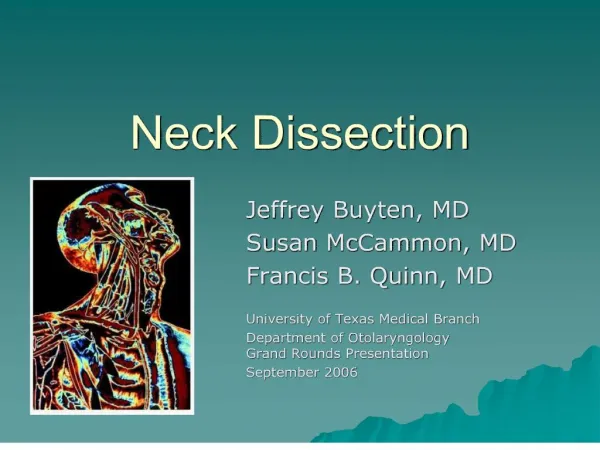
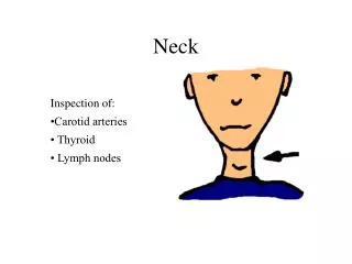
Neck
Neck. Inspection of: •Carotid arteries • Thyroid • Lymph nodes. 54. Neck Inspection. Inspect the neck for symmetry, masses, pulsations, midline location of the trachea, thyromegally, and note the use of accessory muscles of respiration. The Carotid Arteries.

Neck
E N D
Presentation Transcript
Neck Inspection of: •Carotid arteries • Thyroid • Lymph nodes
54. Neck Inspection Inspect the neck for symmetry, masses, pulsations, midline location of the trachea, thyromegally, and note the use of accessory muscles of respiration
The Carotid Arteries The carotid arteries may be palpated medial to the sternocleidomastoids and the trachea. The carotid arteries bifurcate superiorly near the angle of the jaw. The carotid body is found at the bifercation
57, 58. Auscultation of the Carotid Arteries • The bell or diaphragm may be used, the bell may be easier • Auscultate the carotid arteries in both the inferior and superior regions.
55, 56. Palpation of the carotid arteries • Place your fingers on the trachea and slide them laterally towards the medical aspect of the sternocleidomastoid • (you may also use your thumbs) • Palpate low in the neck avoiding the carotid body. Stimulation of the carotid body may activate • the vagus nerve and cause bradycardia and hypotension. Do not palpate both carotids at the same time
Location of Thyroid Gland Figure 11-5. p. 271. Slide 11-5 • The thyroid lies inferior to the cricoid cartilage • The isthus may be palpable in the midline over the trachea below the circoid. • The lobes of the thyroid curve posteriorly around the sides of the trachea • The lobes are covered by thin straplike muscles and the sternocleidomastoides. • These muscles need to be displaced in order to palpate the lodes
Posterior Approach 59. Palpate Thyroid: Anterior Approach
THYROID Palpation - may be done anterior or posterior: • Have the patient extend their chin • Inspect the neck to see if the thyroid is visible • Palpate the cricoid cartilage • Slide your fingers down the trachea to palpate the isthmus • The isthmus is palpated by having your fingers of both hands almost touch in the midline • just below the cricoid membrane and asking the patient to swallow. • Swallowing may be aided by giving the patient some water and asking them to hold it • in their mouth until you ask them to swallow. • Next palpate the lobes • Slide your fingers laterally beneath the sternoceidomastoids. • Note that the lobes of the thyroid run both superiorly and inferiorly to the isthmus • Palpation of the lobes may be aided by slightly displacing the opposite side of the trachea (see the arrows in the diagrams to the right) • Once more ask the patient to swallow during palpation • Note the size, shape, and consistency of the gland. Note any nodules or tenderness
Lymph Nodes of the Head and Neck • Preauricular • Posterior auricular • Occipital • Tonsillar • Submndibular • Submental • Posterior cervical • Superficial cervical • Deep cervical • Supraclavicular
Location of Lymph Nodes •Periauricular (in front of the ear) • Posterior auricular (behind the ear) • Occipital (base of skull) • Tonsillar (jugulodgastric)(angle of jaw) • Submaxillary (mid-jaw) Figure 11-6. p. 271.
Periauricular are located in front of the ear. • Posterior auricular are located behind the ear (not shown in image)
70. Superficial cervical are on the top of the sternomastoid muscle.