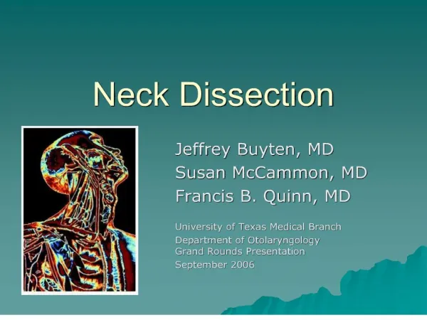
CT NECK
CT NECK . MANDIBLE. LT. MASSETER MUSCLE. MASSETER MUSCLE. SCAN LEVEL. PTERYGOID MUSCLES. PAROTID GLAND. LT. SUBMANDIBULAR GLAND. SCAN LEVEL. EPIGLOTTIS. STERNOCLEIOMASTOID MUSCLE. SUBCUTANEOUS FAT. LT. HYOID BONE. SCAN LEVEL. VALLECULA. PRYIFORM SINUS.
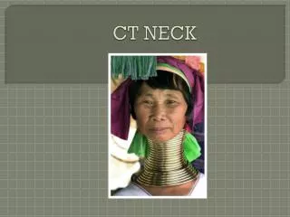

CT NECK
E N D
Presentation Transcript
MANDIBLE LT MASSETER MUSCLE MASSETER MUSCLE SCAN LEVEL PTERYGOID MUSCLES PAROTID GLAND
LT SUBMANDIBULAR GLAND SCAN LEVEL EPIGLOTTIS STERNOCLEIOMASTOID MUSCLE SUBCUTANEOUS FAT
LT HYOID BONE SCAN LEVEL VALLECULA PRYIFORM SINUS JUGULAR VEIN JUGULAR VEIN COMMON CAROTID ARTERIES
LT STERNOCLEIDOMASTOID MUSCLE SCAN LEVEL THYROID CARTILAGE VOCAL CORD
LT THYROID CARTILAGE SCAN LEVEL COMMON CAROTID ARTERY CRICOID CARTILAGE JUGULAR VEIN
LT SCAN LEVEL THYROID GLAND CLAVICLE CLAVICLE FAT FAT ESOPHAGUS TRACHEA
CT scan shows left thyroid mass at the level of the clavicles displacing the trachea to the right.
CT angio neck for carotid CT angiography showed left carotid stenosis
THANK You