Download

1 / 32
320 likes | 725 Vues
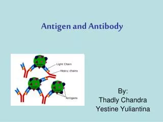
Antigen-Antibody Interactions. Serology - in vitro demonstration of Ag/Ab reaction Ag/Ab reaction = reversible 1. Primary interactions 1) Electrostatic interactions 2) Hydrogen bond 3) Hydrophobic interactions 4) van de Waals force 2. Secondary interactions Precipitation

E N D
Serology - in vitro demonstration of Ag/Ab reaction Ag/Ab reaction = reversible 1.Primary interactions 1) Electrostatic interactions 2) Hydrogen bond 3) Hydrophobic interactions 4) van de Waals force 2.Secondary interactions Precipitation Agglutination Complement activation reactions
Association constant Ab + H AbH (binding of hapten) K = [AbH]/[Ab][H] (association constant between reactants) Affinity = binding of Ab with epitope characterized by association constant Avidity = binding energy between Ab & multivalent Ag IgM > IgG
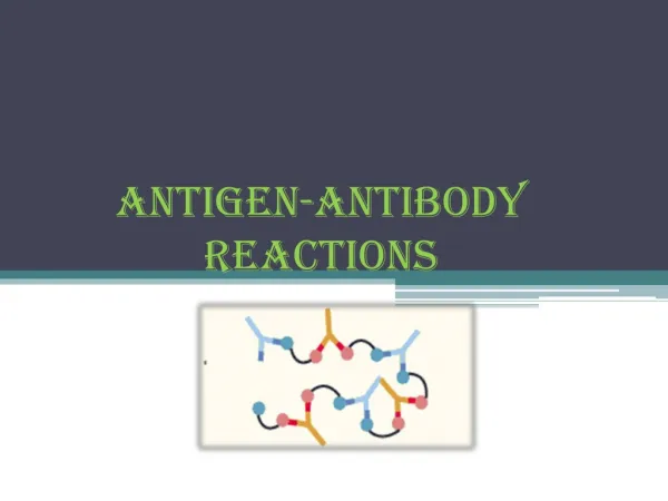
Figure 5.1 Reactions between antibody or antibody fragments and antigens or hapten.
Figure 5.2 A representation of the anti-immunoglobulin (Coombs) test.
Agglutination inhibition (Early home pregnancy test kit)
Figure 5.3 A representation of the precipitin reaction.
Figure 5.4 Gel diffusion by antibodies and a single antigen (A) and antibodies to antigens 1, 2, 3, and their respective antigens (B).
Figure 5.5 Double gel-diffusion patterns showing pattern of identity (left), pattern of nonidentity (center), and pattern of partial identity (right).
Figure 5.6 Radial diffusion, A, B, C, D, and E represent known concentrations of antigen; F and G represent unknown concentrations that can be determined from the graph.
Figure 5.7 Patterns of immunoelectrophoresis of serum proteins.
Figure 5.8 Western blots of serum samples from two HIV-infected individuals and one control subject.
Figure 5.9 Amount of label bound to antibody after incubation of constant amounts of antibody and labeled antigen.
Figure 5.10 Radioimmunoassay, based on the competition of nonlabeled and labeled antigens for antibody.
Solid phase radioimmunoassay (RIA) to detect hepatitis B virus in blood serum
Figure 5.11 A representative ELISA using a well coated directly with antigen
Figure 5.12 A schematic representation of a fluorescence-activated cell sorter (FACS).
Figure 5.13 A schematic representation of the production of monoclonal antibodies.
Figure 5.14 A general procedure for producing transgenic mice.