Download
1 / 59
950 likes | 2.78k Vues
Antigen-antibody reactions. Let’s start. The immune system is a system of biological structures and processes within an organism that protects against disease.

E N D
Let’s start • The immune system is a system of biological structures and processes within an organism that protects against disease. • To function properly, an immune system must detect a wide variety of agents, from viruses to parasitic worms, and distinguish them from the organism’s own healthy tissue.
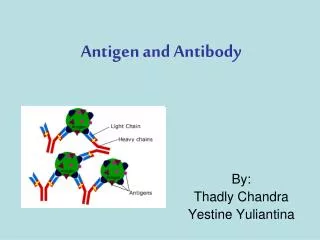
Antigens • Antigens are any substances that are capable, under appropriate conditions, of inducing the formation of antibodies and reacting specifically with the antibodies so produced. • These antigenic molecules may have several antigenic determinants, called epitopes, and each epitope can bind with a specific antibody. Thus, a single antigen can bind to many different antibodies with different binding sites
Chemical Nature of Antigens ((Immunogens A. Proteins The vast majority of immunogens are proteins. These may be pure proteins or they may be glycoproteins or lipoproteins. In general, proteins are usually very good immunogens. B. Polysaccharides Pure polysaccharides and lipopolysaccharides are good immunogens. C. Nucleic Acids Nucleic acids are usually poorly immunogenic. However, they may become immunogenic when single stranded or when complexed with proteins. D. Lipids In general lipids are non-immunogenic, although they may be haptens.
Antibodies • An antibody is a protein produced by the body’s immune cells “ activated B-lymphocytes” when it detects a foreign antigen. • Classes of antibodies: • IgG: The main antibody in blood(70%), made in 2ry immune response and has a long half life”up to 20 years”. • IgM: accounts for(10%) of Igs, has a key role in 1ry immune response. • IgA: (20%) of Igs present in serum and secretions. • IgE: (0.001%), involved in allergy and parasitic infections. • IgD: (1%) present on the surface of B cells, so that it plays a role in induction of Ab production
Nature of antigen-antibody reactions • A. Lock and Key Concept The combining site of an antibody is located in the Fab portion of the molecule and is constructed from the hypervariable regions of the heavy and light chains • B. Non-covalent Bonds The bonds that hold the antigen to the antibody combining site are all non-covalent in nature. These include hydrogen bonds, electrostatic bonds, Van der Waals forces and hydrophobic bonds. • C. ReversibilitySince antigen-antibody reactions occur via non-covalent bonds, they are by their nature reversible.
Affinity and avidity • AffinityAntibody affinity is the strength of the reaction between a single antigenic determinant and a single combining site on the antibody. • AvidityAvidity is a measure of the overall strength of binding of an antigen with many antigenic determinants and multivalent antibodies.
Specificity and cross reactivity Specificity Specificity refers to the ability of an individual antibody combining site to react with only one antigenic Cross reactivity Cross reactivity refers to the ability of an individual antibody combining site to react with more than one antigenic determinant Sensitivity Cross reactivity refers to the ability of an individual antibody to locate antigen even when it is greatly diluted.
Factors affecting measurement of antigen-antibody reactions • The only way that one knows that an antigen-antibody reaction has occurred is to have some means of directly or indirectly detecting the complexes formed between the antigen and antibody. • The ease with which one can detect antigen-antibody reactions will depend on a number of factors. 1. AffinityThe higher the affinity of the antibody for the antigen, the more stable will be the interaction. Thus, the ease with which one can detect the interaction is enhanced. 2. Avidity Reactions between multivalent antigens and multivalent antibodies are more stable and thus easier to detect.
Factors affecting measurement of antigen-antibody reactions 3. Antigen to antibody ratio The ratio between the antigen and antibody influences the detection of antigen-antibody complexes because the size of the complexes formed is related to the concentration of the antigen and antibody. 4. Physical form of the antigenThe physical form of the antigen influences how one detects its reaction with an antibody. If the antigen is a particulate, one generally looks for agglutination of the antigen by the antibody. If the antigen is soluble one generally looks for the precipitation of the antigen after the production of large insoluble antigen-antibody complexes.
Types of Antigen – Antibody Reaction • The types of antigen – antibody reactions are: • Precipitation Reaction. • Agglutination Reaction. • Complement Fixation. • ELISA – Enzyme Linked ImmunoSorbentAssay • Immunofluorescence.
Agglutination • Agglutination is the clumping of particles. • Agglutination occurs if an antigen is mixed with its corresponding antibody called isoagglutinin. • This term is commonly used in blood grouping.
Agglutination This occurs in biology in several examples: • The clumping of cells such as bacteria or red blood cells in the presence of an antibody or complement. The antibody or other molecule binds multiple particles and joins them, creating a large complex. This increases the efficacy of microbial elimination by phagocytosis as large clumps of bacteria can be eliminated in one pass, versus the elimination of single microbial antigens. • Another example occurs when people are given blood transfusions of the wrong blood group.
Characteristics • Is the aggregation of particulate matter due to combination with specific antibody. • Takes place on the surface of the particle and thus antigen must be exposed and able to bind with antibody • Antigens may be: • On a cell (direct agglutination) • Attached to latex spheres (indirect or passive agglutination) • Agglutination reaction is aided by elevated temperature (37-56°C) and by movement which increases the contact between antigen and antibody. • Clear supernatant. • Clumps aggregate and settle as large visible clumps.
Steps in Agglutination 1- Sensitization Involves antigen-antibody combination through single antigenic determinants on the particle surface Antibody molecules attach to their corresponding Antigenic site (epitope) on the red blood cell membrane. There is no visible clumping.
2- Aggregative Stage (lattice formation) • Represents the sum of interaction between antibody and multiple antigenic determinants on a particle • Dependent on environmental conditions as well as the relative concentrations of antigen and antibody Antibody molecules crosslink RBCs forming a lattice that results in visible clumping or agglutination.
Uses of Agglutination Reactions • Aid in the identification, by means of known antisera (serum containing antibodies specific for a given antigen), microorganisms cultured from clinical specimens. • Help estimate the titer of antibacterial agglutinins in the serum of patients with unknown diseases.
Types of Agglutination Reactions • Direct Agglutination • Passive Agglutination • Reverse Passive Agglutination • Agglutination inhibition • Hemagglutination-inhibition • Coagglutination/Conglutination
Direct agglutination • Occurs when antigens are found naturally on a particle (Serotyping of Salmonella) • e.g. identification of bacterial types , O antigen (somatic) - compact, fine and granular agglutination , H antigen (flagellar) - form a loosely woven network of clumped cells (coarse and floccular), called snowflake agglutination
Hemagglutination • Hemagglutination, is agglutination that involves red blood cells (RBCs). It has two common uses in the laboratory: • Blood typing • Quantification of virus dilutions in a Haemagglutination assay.
Blood typing • Blood type can be determined by usingantibodies that bind to the A or B blood group antigens in a sample of blood. • For example, if antibodies that bind the A blood group are added and agglutination occurs, the blood is either type A or type AB. • To determine between type A or type AB, antibodies that bind the B group are added and if agglutination does not occur, the blood is type A. • If agglutination does not occur with either antibodies that bind to type A or type B antigens, then neither antigen is present on the blood cells, which means the blood is type O.
Viral hemagglutination assay • Hemagglutination phenomenon is almost commonly used for diagnosis of infection produced by some viruses. • The presence of virus in infected cell cultures can be detected by hemagglutination • The identity of the virus or of antibodies in a patient’s serum can be determined by specific inhibition of that hemagglutination.
Viral hemagglutination assay • The basis of the HAI assay is that antibodies to that particular virus (for example-influenza virus) will prevent attachment of the virus to RBC. Therefore hemagglutination is inhibited when antibodies are present.
Passive agglutination • Employs particles that are coated with antigens not normally found on their surfaces. • Inert materials commonly used: • Bentonite • Colloidion • Latex particles • Colloidal charcoal • Passive agglutination tests have been used to detect rheumatoid factor and antinuclear antibody.
Reverse Passive agglutination • Reverse Passive agglutination - antibody rather than antigen is attached to a carrier particle. • Several kits are available today for rapid identification of such antigens from such infectious agents as group A and B streptococci, Staph. , Neisseria, and others.
Agglutination inhibition • Agglutination inhibition - based on competition between particulate and soluble antigens for limited antibody combining sites, and a lack of agglutination is an indicator of a positive reaction.
The classic example of agglutination inhibition is the early types of home pregnancy test kits included latex particles coated with human chorionic gonadotropin (HCG) and antibody to HCG • The addition of urine from a pregnant woman, which contained HCG, inhibited agglutination of the latex particles when the anti-HCG antibody was added; thus the absence of agglutination indicated pregnancy.
Coagglutination/Conglutination • Coagglutination/Conglutination - name given to systems using bacteria as inert particles to which antibody is attached (S. aureus). • The Fc region of antibody attaches to protein A of staphylococcal cell leaving the Fab region to combine with the antigen • Killed staphylococcal cells coated with antibody can be used to identify bacteria and detect soluble extracellular bacterial antigens in specimens and body fluids.
Antiglobulin mediated agglutination • The antiglobulin test (Coomb’s test) can be used to detect red cells sensitized with IgG antibodies • In order for agglutination to occur an additional of anti-antibody , which reacts with the Fc portion of the IgG antibody • This will form a "bridge" between the antibodies or complement coating the red cells, causing agglutination.
Instrumentation • Several systems were developed to increase sensitivity of results reading, many of these utilize turbidimetry • Particle Enhanced Turbidimetric Inhibition Assay (PETINA) • Particle counting immunoassay (PACIA)
Particle Enhanced Turbidimetric Inhibition Assay (PETINA) • This method uses the creation of light scattering particles to measure drug levels. • The latex particle-bound drug binds to the drug-specific antibody, forming insoluble light- scattering aggregates. • This causes an increase in the turbidity of the reaction mixture. • Non-particle- bound drug in the patient sample competes with the particle-bound drug for antibody binding sites, inhibiting the formation of insoluble aggregates. • Therefore the rate of increase of absorbance (hence the rate of the increase in turbidity,) is inversely proportional to the concentration of the drug.
Particle counting immunoassay (PACIA) • Involves measurement of the number of residual non-agglutinating particles in a specimen. • Latex particles are coated with whole antibody molecule, if antigen is present complexes will form and will screened out by counter . • An inverse relationship exists between the number of unagglutinated particles counted and the amount of unknown in the patient specimen.
Qualitative Agglutination Test • Agglutination tests can be used in a qualitative manner to assay for the presence of an antigen or an antibody. • The antibody is mixed with the particulate antigen and a positive test is indicated by the agglutination of the particulate antigen.
Quantitative Agglutination Test • Agglutination tests can also be used to quantitate the level of antibodies to particulate antigens. • In this test • one makes serial dilutions of a sample to be tested for antibody • and then adds a fixed number of red blood cells or bacteria or other such particulate antigen • and determines the maximum dilution, which gives agglutination. • The maximum dilution that gives visible agglutination is called the titer. • The results are reported as the reciprocal of the maximal dilution that gives visible agglutination. This can be done using a microtiter plate.
Quantitative Agglutination Test 1/1024 1/256 1/512 1/128 1/16 1/64 1/32 Pos. 1/8 Neg. 1/4 1/2 Titer Patient 64 1 8 2 512 3 <2 4 32 5 128 6 32 7 4 8
Determining Antibody titer • Titer is the quantity of a substance required to produce a reaction with a given volume of another substance. • Antibody titer is the highest dilution of the biological sample that still results in agglutination, with no agglutination being observed at any higher dilution. • The term is used in serological reactions and is determined by preparing serial dilutions of antibody to which a constant amount of antigen is added.
Advantages and disadvantages Advantages of agglutination tests: • Low individual test cost. • Ability to obtain semi quantitative results. • Short time to obtain result. • Don’t need expensive instrument. • Agglutination of insoluble native antigens or antigen-coated particles simple to read with or without the aid of a microscope • Increased degree of sensitivity • Great variety of detectable substances • If the sample contain micro-organisms, it does not need to be viable
Agglutination Requirements • Availability of stable cell or particle suspension • Presence of one or more antigens close to the surface • Knowledge that ‘incomplete’ or nonagglutinating antibodies are not detectable without modifications, e.g. antiglobulin (Coomb’s) technic
Advantages and disadvantages Disadvantages of agglutination tests: • It must kept in mind that agglutination reaction are screening tests only, and negative result doesn't rule out disease; the quantity of antigen or antibody may be below the sensitivity of the test system • May give false positive or negative results
False positive results in agglutination reactions • Overcentrifugation • Button is packed too tight and is difficult to resuspend. • Contaminated Equipment • Dust, dirt or fingerprints may cause cells to clump. • Autoagglutination • Test cells clump without specific AB present, mainly problematic with RBCs. • Delay in Reading Tests • Dried out Ag may look like agglutination • Saline Stored in Glass Bottles • Colloidal Silica may leach out and cause agglutination • Using plasma instead of serum, hemolysed or lipemicsample. • Cross reaction
False negative • Undercentrifugation Cells may not be close enough to interact • Inactive Reagents May cause improper storage • Incorrect Incubation Temperature May result in the lack of association for AGs and ABs • Insufficient Incubation Time AGs and ABs may not have time for association • ProzonePhenomenon Too much patient AB for amount of testing • Delay in Testing Procedure AB may be eluted from RBCs
Experiment 1 Determination of the titer of blood group cold antibody