Download
1 / 33
360 likes | 1.02k Vues
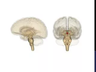
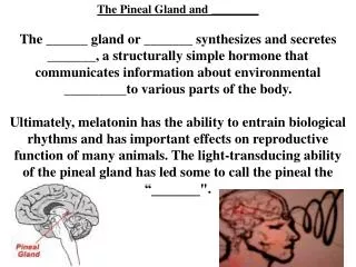
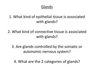
Glands 1. What kind of epithelial tissue is associated with glands? 2. What kind of connective tissue is associated with glands? 3. Are glands controlled by the somatic or autonomic nervous system? 4. What are the 2 categories of glands?. Ductless Glands Diffuse tissue. Pineal gland.

E N D
Glands1. What kind of epithelial tissue is associated with glands?2. What kind of connective tissue is associated with glands?3. Are glands controlled by the somatic or autonomic nervous system?4. What are the 2 categories of glands?
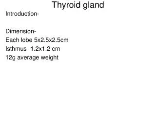
Ductless Glands Diffuse tissue Pineal gland Hypothalamus Pituitary gland Thyroid gland Parathyroid glands (on dorsal aspect of thyroid gland) Thymus Adrenal glands Pancreas Ovary (female) Testis (male) 592
Signaling MoleculesChemicals produced by the body that target cells, causing them to change their behavior.Ex. Parathyroid hormone targeting osteoclastsInclude: Hormones and Neurotransmitters
FunctionsInternal Control-Coordinate body activities (gametes)-Maintain homeostasis (calcium)-Growth and development-Regulate metabolism-Helps body respond to stress (adrenaline)Works with ANS (Endocrine and ANS are 2 primary systems that regulate the human body)-ANS controls release of hormones-Hormones can initiate or inhibit nerve impulses-Endocrine system slower, but longer lasting
Amino Acid Based Hormones-Proteins -Most common -Bind to surface receptors (G protein linked)-Amines -Small, simple, derived from a.a. tyrosine -Most act like protein hormones and bind to surface receptors. -Include: epinephrine, norepinephrine, melatonin -NOTE: amines produced by the thyroid gland will act more like steroid hormones instead of proteins.
Protein hormones use G protein linked receptors (2nd messenger Systems) Extracellular fluid 1 Hormone (1st messenger) binds receptor. Adenylate cyclase G protein (GS) 5 cAMP acti- vates protein kinases. Receptor Active protein kinase GDP Inactive protein kinase 2 3 4 Receptor activates G protein (GS) G protein activates adenylate cyclase. Adenylate cyclase converts ATP to cAMP (2nd messenger). Hormones that act via cAMP mechanisms: Epinephrine ACTH FSH LH Triggers responses of target cell (activates enzymes, stimulates cellular secretion, opens ion channel, etc.) Glucagon PTH TSH Calcitonin G protein can be activated or inhibited depending on the hormone. Cytoplasm 594
Fat soluble (lipophilic) hormones: Steroids and thyroid amines Steroids -Cholesterol -Ovary -Testes -Cortex of adrenal gland Steroid hormone Plasma membrane Extracellular fluid 1 The steroid hormone diffuses through the plasma membrane and binds an intracellular receptor. Cytoplasm Receptor protein Receptor- hormone complex 2 The receptor- hormone complex enters the nucleus. Hormone response elements Nucleus The receptor- hormone complex binds a hormone response element (a specific DNA sequence). 3 DNA 4 Binding initiates transcription of the gene to mRNA. mRNA *Thyroid amine receptor is always bound to the DNA The mRNA directs protein synthesis. 5 New protein 599
Eicosanoids: Hormone-Like; Locally acting (No circulation); Paracrine vs. Autocrine -made of fatty acids from phospholipids -released due to mechanical or chemical stimulation of cell membranes (Except RBC’s) -Bind to surface receptors (G protein linked) -Rapidly inactivated -Production inhibited by aspirin and ibuprofen Prostaglandins -Influence smooth muscle, pain, inflammation Leukotrienes -Respiratory system -Inflammation and bronchoconstriction
Target Cell Specificity (Activation)Hormones bind to a matching receptor, but their activation of the target cell depends on: • Blood levels of hormone • Number of receptors • Up regulation • Down regulation (drug tolerance) • Affinity (binding strength high or low)
Cells decrease numbers of receptors in response to higher levels of a hormone. Cells increase numbers of receptors in response to low levels of a hormone.
Epinephrine has better binding affinity with alpha receptors so therefore will cause a greater response at cells with alpha receptors.
Interaction of Hormones at Target Cells • Permissiveness: One hormone cannot exert it’s full effect without another hormone present. (Thyroxine and adrenaline at the heart) • Synergism: 2 or more hormones with the same effects at target cell can work together to amplify their effect. • Antagonism: One hormone opposing the action of another hormone. (insulin vs. glucagon and blood sugar)
Posterior pituitary, Adrenal , Pineal gland Nutrients and ions (a) Humoral Stimulus (b) Neural Stimulus (c) Hormonal Stimulus Preganglionic sympathetic fibers stimulate adrenal medulla cells... 1 1 1 Capillary blood contains low concentration of Ca2+, which stimulates... The hypothalamus secretes hormones that... Hypothalamus receptors CNS (spinal cord) Capillary (low Ca2+ in blood) 2 …stimulate the anterior pituitary gland to secrete hormones that… Thyroid gland (posterior view) Parathyroid glands Pituitary gland Preganglionic sympathetic fibers Thyroid gland Adrenal cortex Gonad (Testis) Medulla of adrenal gland Parathyroid glands PTH Capillary …secretion of parathyroid hormone (PTH) by parathyroid glands* 2 …to secrete catechola- mines (epinephrine and norepinephrine) 2 …stimulate other endocrine glands to secrete hormones 3 Hypothalamus 601
Negative Feedback (Stabilizing)Blood Glucose Pancreas Insulin SecretedBlood glucose Increase cell intake of glucose
Positive Feedback (Disruptive)Mother produces milkBaby sucklesStimulates mom to make prolactinMore milk productionMore sucklingMore milkEtc.
Figure 16.5 The hypothalamus controls release of hormones from the pituitary gland in two different ways. (2 of 2) Aka: Adenohypophysis Anterior Pituitary: Lots of receptors Hypothalamus Hypothalamic neurons synthesize GHRH, GHIH, TRH, CRH, GnRH, PIH. Anterior lobe of pituitary Superior hypophyseal artery 1 When appropriately stimulated, hypothalamic neurons secrete releasing or inhibiting hormones into the primary capillary plexus. 2 Hypothalamic hormones travel through portal veins to the anterior pituitary where they stimulate or inhibit release of hormones made in the anterior pituitary. Hypophyseal portal system • Primary capillary plexus A portal system is two capillary plexuses (beds) connected by veins. 3 In response to releasing hormones, the anterior pituitary secretes hormones into the secondary capillary plexus. This in turn empties into the general circulation. • Hypophyseal portal veins • Secondary capillary plexus GH, TSH, ACTH, FSH, LH, PRL Anterior lobe of pituitary glandular 601 © 2013 Pearson Education, Inc.
Figure 16.5 The hypothalamus controls release of hormones from the pituitary gland in two different ways. (1 of 2) Aka: Neurohypophysis Posterior Pituitary: Paraventricular nucleus Hypothalamus 1 Hypothalamic neurons synthesize oxytocin or antidiuretic hormone (ADH). Posterior lobe of pituitary Optic chiasma Supraoptic nucleus Infundibulum (connecting stalk) 2 Oxytocin and ADH are transported down the axons of the hypothalamic- hypophyseal tract to the posterior pituitary. Inferior hypophyseal artery Hypothalamic- hypophyseal tract Axon terminals 3 Oxytocin and ADH are stored in axon terminals in the posterior pituitary. Posterior lobe of pituitary 4 When hypothalamic neurons fire, action potentials arriving at the axon terminals cause oxytocin or ADH to be released into the blood. Oxytocin ADH Neurohormones © 2013 Pearson Education, Inc. 600
Figure 16.17 Stress and the adrenal gland. Short-term stress Prolonged stress Stress Adrenaline (epinephrine) Noradrenaline (norepinephrine) Nerve impulses Hypothalamus CRH (corticotropin- releasing hormone) Spinal cord Corticotropic cells of anterior pituitary Preganglionic sympathetic fibers To target in blood Adrenal cortex (secretes steroid hormones) Adrenal medulla (secretes amino acid– based hormones) ACTH Catecholamines (epinephrine and norepinephrine) Mineralocorticoids Glucocorticoids Long-term stress response Short-term stress response • Heart rate increases • Kidneys retain sodium and water • Proteins and fats converted to glucose or broken down for energy • Blood pressure increases • Bronchioles dilate • Blood volume and blood pressure rise • Liver converts glycogen to glucose and releases glucose to blood • Blood glucose increases • Immune system supressed • Blood flow changes, reducing digestive system activity and urine output • Metabolic rate increases © 2013 Pearson Education, Inc. 617
Neural Hormonal inhibits
Elimination of HormonesSteroid hormones broken down by cell.Hormones in circulation filtered by liver and kidneysHalf life variesvideo
Figure 16.13 Effects of parathyroid hormone on bone, the kidneys, and the intestine. Hypocalcemia (low blood Ca2+) PTH release from parathyroid gland Activation of vitamin D by kidney Ca2+ reabsorption in kidney tubule Osteoclast activity in bone causes Ca2+ and PO43- release into blood Ca2+ absorption from food in small intestine Ca2+ in blood Initial stimulus Physiological response © 2013 Pearson Education, Inc. 611 Result
Figure 16.12a The parathyroid glands. Pharynx (posterior aspect) Thyroid gland Parathyroid glands Esophagus Trachea 610 © 2013 Pearson Education, Inc.
Calcitonin • Produced by Thyroid Gland • No known physiological role in humans • Antagonist to parathyroid hormone (PTH) • At higher than normal doses • Inhibits osteoclast activity and release of Ca2+ from bone matrix • Stimulates Ca2+ uptake and incorporation into bone matrix © 2013 Pearson Education, Inc.
Figure 16.14a Microscopic structure of the adrenal gland. Capsule Zona glomerulosa Zona fasciculata Adrenal gland Neural • Medulla Cortex • Cortex Glandular Kidney Zona reticularis (Corticosteroids) -mineralcorticoids -glucocorticoids -gonadocorticoids Adrenal medulla Medulla Drawing of the histology of the adrenal cortex and a portion of the adrenal medulla 612 © 2013 Pearson Education, Inc.
Glucocorticoids: Cortisol • Released in response to ACTH, patterns of eating and activity (biological clock), and stress • Helps body resist stress—formation of glucose from fats and proteins • Promotes rises in blood glucose, fatty acids, and amino acids • Increases blood pressure to quickly distribute nutrients to cells © 2013 Pearson Education, Inc.
Excessive levels of glucocorticoids:-Depress cartilage and bone formation-Inhibit inflammation-Depress the immune system-Glucocorticoid drugs (corticosteroids) are given to help with chronic inflammatory disorders (rheumatoid arthritis, dermatitis and autoimmune diseases)
Figure 16.17 Stress and the adrenal gland. Short-term stress Prolonged stress Stress Biological Clock Nerve impulses Hypothalamus CRH (corticotropin- releasing hormone) Spinal cord Corticotropic cells of anterior pituitary Preganglionic sympathetic fibers To target in blood Adrenal cortex (secretes steroid hormones) Adrenal medulla (secretes amino acid– based hormones) ACTH (Cortisol) Catecholamines (epinephrine and norepinephrine) Mineralocorticoids Glucocorticoids Long-term stress response Short-term stress response • Heart rate increases • Kidneys retain sodium and water • Proteins and fats converted to glucose or broken down for energy • Blood pressure increases • Bronchioles dilate • Blood volume and blood pressure rise • Liver converts glycogen to glucose and releases glucose to blood • Blood glucose increases • Immune system supressed (Anti-inflammatory effects) • Blood flow changes, reducing digestive system activity and urine output • Metabolic rate increases © 2013 Pearson Education, Inc. 617
Imbalances of Glucocorticoids • Hypersecretion—Cushing‘s disease • Depresses cartilage and bone formation • Inhibits inflammation • Depresses immune system • Disrupts cardiovascular, neural, and gastrointestinal function • Hyposecretion—Addison's disease - Decrease in glucose and Na+ levels - Weight loss, severe dehydration, and hypotension © 2013 Pearson Education, Inc.
Figure 16.16 The effects of excess glucocorticoid. Patient before onset. Same patient with Cushing’s syndrome. The white arrow shows the characteristic “buffalo hump” of fat on the upper back. 615 © 2013 Pearson Education, Inc.
Gonadocorticoids (Sex Hormones) • Most weak androgens (male sex hormones) converted to testosterone in tissue cells, some to estrogens • May contribute to • Onset of puberty • Appearance of secondary sex characteristics • Sex drive in women • Estrogens in postmenopausal women © 2013 Pearson Education, Inc.