Download
1 / 99
1.01k likes | 1.29k Vues
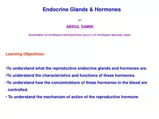
Physiologic anatomical peculiarities of endocrine system in children. Methodics of endocrine glands investigation. Semiotics of hypo- and hyperfunction of some endocrine glands and diseases of the endocrine system. By Nykytyuk S. Major endocrine glands. ( Male left, female on the right.)

E N D
Physiologic anatomical peculiarities of endocrine system in children. Methodics of endocrine glands investigation. Semiotics of hypo- and hyperfunction of some endocrine glands and diseases of the endocrine system.By Nykytyuk S
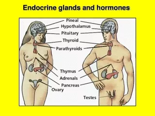
Major endocrine glands. (Male left, female on the right.) 1.Pineal gland 2.Pituitary gland 3.Thyroid gland 4.Thymus 5.Adrenal gland 6.Pancreas 7.Ovary 8.Testis
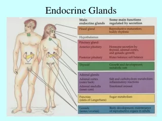
The endocrine system provides a chemical connection from the hypothalamus of the brain to all the organs that control body metabolism, growth and development, and reproduction. • There are two types of hormones secreted in the endocrine system: (1) steroidal and (2) nonsteroidal, or protein based, hormones. The endocrine system regulates its hormones through negative feedback control. Increases in hormone activity decreases the production of that hormone. The immune system and other factors contribute as control factors also, maintaining constant levels of hormones.
Hypothalamus produces Thyrotropin-releasing hormone (TRH) Gonadotropin-releasing hormone (GnRH) Growth hormone-releasing hormone (GHRH) Corticotropin-releasing hormone (CRH) Somatostatin (SS; also GHIH, growth factor-inhibiting hormone) Dopamine (DA) Pineal Gland produces Melatonin Endocrine glands and the hormones secreted
Pituitary gland (hypophysis) produces Anterior pituitary lobe (adenohypophysis) Growth hormone (GH) Prolactin (PRL) Adrenocorticotropic hormone (ACTH, corticotropin) Thyroid-stimulating hormone (TSH, thyrotropin) Follicle-stimulating hormone (FSH, a gonadotropin) Luteinizing hormone (LH, a gonadotropin) Posterior pituitary lobe (neurohypophysis) Oxytocin (ocytocin) Arginine vasopressin (AVP; also ADH, antidiuretic hormone) Lipotropin Endocrine glands and the hormones secreted
Thyroid gland produces Triiodothyronine (T3), the potent form of thyroid hormone Thyroxine (T4), a less active form of thyroid hormone Calcitonin Parathyroid gland produces Parathyroid hormone (PTH) Heart produces Atrial-natriuretic peptide (ANP) Stomach and intestines produce Cholecystokinin (CCK) Gastrin Ghrelin Neuropeptide Y (NPY) Secretin Somatostatin Endocrine glands and the hormones secreted
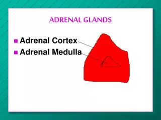
Liver produces Insulin-like growth factor (IGF) Angiotensinogen Thrombopoietin Islets of Langerhans in the pancreas produce Insulin Glucagon Somatostatin Adrenal glands produce Adrenal cortex Glucocorticoids (chiefly cortisol) Mineralocorticoids (chiefly aldosterone) Androgens (including DHEA and testosterone) Adrenal medulla Adrenaline (epinephrine) Noradrenaline (norepinephrine) Testosterone Endocrine glands and the hormones secreted
Endocrine glands and the hormones secreted • Kidney produces • Renin • Erythropoietin (EPO) • Calcitriol (the active form of vitamin D3) • Skin produces • Vitamin D3 (calciferol) • Adipose tissue • Leptin • Estrogens (mainly estrone
In males only Testes Androgens (chiefly testosterone) In females only Ovarian follicle Estrogens (mainly estradiol) Corpus luteum Progesterone Estrogens (mainly estradiol) Placenta (when pregnant) Progesterone Estrogens (mainly estriol) Human chorionic gonadotropin (HCG) Human placental lactogen (HPL) Endocrine glands and the hormones secreted
The pineal gland is a reddish-gray body about the size of a pea (8 mm in humans), located just rostro-dorsal to the superior colliculus and behind and beneath the stria medullaris, between the laterally positioned thalamic bodies. It is part of the epithalamus. Pineal gland
The pineal gland is large in children, but shrinks at puberty. It appears to play a major role in sexual development, hibernation in animals, metabolism, and seasonal breeding. The abundant melatonin levels in children is believed to inhibit sexual development, and pineal tumors have been linked with precocious puberty. When puberty arrives, melatonin production is reduced. Calcification of the pineal gland is typical in adults.
The pituitary gland, or hypophysis, is an endocrine gland about the size of a pea that sits in a small, bony cavity (sella turcica) at the base of the brain. The pituitary gland secretes hormones regulating homeostasis, including trophic hormones that stimulate other endocrine glands. It is functionally connected to the hypothalamus by the median eminence. Pituitary gland
Posterior pituitary (neurohypophysis) • The posterior lobe is connected to a part of the brain called the hypothalamus via the infundibulum (or stalk), giving rise to the tuberoinfundibular pathway. Hormones are made in nerve cell bodies positioned in the hypothalamus, and these hormones are then transported down the nerve cell's axons to the posterior pituitary. Hypothalamic neurons fire such hormones, releasing them into the capillaries of the pituitary gland. • The hormones secreted by the posterior pituitary are • Oxytocin comes from the paraventricular nucleus in the Hypothalamus • Antidiuretic hormone (ADH - also known as vasopressin), comes from the supraoptic nucleus in the Hypothalamus
The anterior pituitary produces and secretes: growth hormone prolactin follicle-stimulating hormone luteinizing hormone thyroid-stimulating hormone adrenocorticotropic hormone endorphins and other hormones It does this in response to releasing hormones produced by the hypothalamus. These travel to the anterior lobe by way of a special capillary system, called the hypothalamic-hypophyseal portal system. These hypothalamic signalling hormones include: TRH (thyrotropin-releasing hormone) CRH (corticotropin-releasing hormone) DA (dopamine, "prolactin inhibiting factor"/PIF) GnRH (gonadotropin-releasing hormone) GHRH (growth hormone releasing hormone) Anterior pituitary (Adenohypophysis)
Intermediate lobe • In adult humans it is just a thin layer of cells between the anterior and posterior pituitary, nearly indistinguishable from the anterior lobe. The intermediate lobe produces melanocyte-stimulating hormone (MSH), although this function is often (imprecisely) attributed to the anterior pituitary.
Functions The pituitary gland helps control the following body processes: • Growth • Blood pressure • Some aspects of pregnancy and childbirth • Breast milk production • Sex organ functions in both women and men • Thyroid gland function • The conversion of food into energy (metabolism) • Water and osmolarity regulation in the body
Adrenocorticotropic hormone • ACTH acts through the stimulation of cell surface ACTH receptors, which are primarily located on the adrenocortical cells. ACTH stimulates the cortex of the adrenal gland and boosts the synthesis of corticosteroids, mainly glucocorticoids but also mineralcorticoids and sex steroids (androgens). Together with ACTH the hormones lipotropin, melanocyte-stimulating hormone (MSH), β-endorphin and met-enkephalin are also released. ACTH is also related to the circadian rhythm in many organisms.
Growth hormone (GH or somatotropin) is a polypeptidehormone synthesised and secreted by the anterior pituitary gland which stimulates growth and cell reproduction in humans Growth hormone
Functions of GH • Effects of growth hormone on the tissues of the body can generally be described as anabolic (building up). Like most other protein hormones GH acts by interacting with a specific receptor on the surface of cells. • Height growth in childhood is the best known effect of GH action, and appears to be stimulated by at least two mechanisms. • 1. GH directly stimulates division and multiplication of chondrocytes of cartilage. These are the primary cells in the growing ends (epiphyses) of children's long bones (arms, legs, digits). • 2. GH also stimulates production of insulin-like growth factor 1 (IGF1, formerly known as somatomedin C), a hormone homologous to proinsulin.
Growth hormone excess: (acromegaly and pituitary gigantism) • The most common disease of GH excess is a pituitary tumor comprised of somatotroph cells of the anterior pituitary. These somatotroph adenomas are benign and grow slowly, gradually producing more and more GH. Prolonged GH excess thickens the bones of the jaw, fingers and toes. Resulting heaviness of the jaw and increased thickness of digits is referred to as acromegaly. GH-secreting tumors are typically recognized in the 5th decade of life. It is extremely rare for such a tumor to occur in childhood, but when it does the excessive GH can cause excessive growth, traditionally referred to as pituitary gigantism.
Growth hormone deficiency (GHD) Deficiency of GH produces significantly different problems at various ages. In children, growth failure and short stature are the major manifestations of GH deficiency. In adults the effects of deficiency are more subtle, and may include deficiencies of strength, energy, and bone mass, as well as increased cardiovascular risk.
Other GH uses and treatment indications • Many other conditions besides GH deficiency cause poor growth, but growth benefits (height gains) are often poorer than when GH deficiency is treated. Examples of other causes of shortness often treated with growth hormone are Turner syndrome, chronic renal failure, Prader-Willi syndrome, intrauterine growth retardation, and severe idiopathic short stature. Higher ("pharmacologic") doses are required to produce significant acceleration of growth in these conditions, producing blood levels well above physiologic.
Thyroid-stimulating hormone • Thyroid-stimulating hormone (also known as TSH or thyrotropin) is a hormone synthesized and secreted by thyrotrope cells in the anterior pituitary gland which regulates the endocrine function of the thyroid gland. TSH stimulates the thyroid gland to secrete the hormones thyroxine (T4) and triiodothyronine (T3). TSH production is controlled by a Thyrotropin Releasing Hormone, (TRH), which is manufactured in the hypothalamus and transported to the pituitary gland, where it increases TSH production and release. Somatostatin is also produced by the hypothalamus, and has an opposite effect on the pituitary production of TSH, decreasing or inhibiting its release.
Primarily Abnormal Pituitary Function • Higher than normal levels of TSH combined with high levels of thyroid hormone (T3 and T4) may indicate dysfunction of the hypothalamus and pituitary gland. In these case, a high TSH is often produced by a benign tumor of the pituitary (adenoma). Conversely, low levels of TSH, while blood levels of T3 and T4 are also low, indicates abnormally low function of the pituitary, known as hypopituitarism.
Primarily Abnormal Thyroid function • On the other hand, due to the negative feedback described above, abnormally high levels of Thyroid hormone, due to overproduction in the thyroid, results in low TSH levels. This occurs in diseases such as hyperthyroidism or Grave's disease. Conversely, an underproduction of T3 and T4 caused by diseases such as congenital hypothyroidism (cretinism), hypothyroidism or thyroid hormone resistance, gives rise to an increase in the measured TSH.
Clearly both TSH and T3 and T4 should be measured to ascertain where a specific thyroid disfunction is caused by primary pituitary or by a primary thyroid disease. If both are up (or down) then the problem is probably in the pituitary. If the one component (TSH) is up, and the other (T3 and T4) is down, then the disease is probably in the thyroid itself. The same holds for a low TSH, high T3 and T4 finding.
Prolactin • Prolactin is a peptide hormone synthesised and secreted by lactotrope cells in the adenohypophysis (anterior pituitary gland). It is also produced in other tissues including the breast and the decidua. Pituitary prolactin secretion is regulated by neuroendocrine neurons in the hypothalamus, most importantly by neurosecretory dopamine neurons of the arcuate nucleus, which inhibit prolactin secretion.
Prolactin has many effects, the most important of which is to stimulate the mammary glands to produce milk (lactation). Increased serum concentrations of prolactin during pregnancy cause enlargement of the mammary glands of the breasts and increases the production of milk. However, the high levels of progesterone during pregnancy act directly on the breasts to stop ejection of milk. It is only when the levels of this hormone fall after childbirth that milk ejection is possible.
Follicle-stimulating hormone • Follicle stimulating hormone (FSH) is a hormone synthesised and secreted by gonadotropes in the anterior pituitary gland. In the ovary FSH stimulates the growth of immature Graafian follicles to maturation. As the follicle grows it releases inhibin, which shuts off the FSH production. In men, FSH enhances the production of androgen-binding protein by the Sertoli cells of the testes and is critical for spermatogenesis. FSH and LH act synergistically in reproduction.
High FSH levels • High FSH levels are indicative of situations where the normal restricting feedback from the gonad is absent, leading to an unrestriced pituitary FSH production. While this is typical in the menopause, it is abnormal in the reproductive years. There it may be a sign of: • Premature menopause • Gonadal dysgenesis, Turner syndrome • Castration • Swyer syndrome • Certain forms of CAH • Testicular failure
Deficient FSH activity • Diminished secretion of FSH can result in failure of gonadal function (hypogonadism). This condition is typically manifest in males as failure in production of normal numbers of sperm. In females, cessation of reproductive cycles is commonly observed. Conditions with very low FSH secretions are: • Kallmann syndrome • Hypothalamic suppression • Hypopituitarism • Hyperprolactinemia • Gonadotropin deficiency • Gonadal suppression therapy • GnRH antagonist • GnRH agonist (downregulation)
Luteinizing hormone • Luteinizing hormone (LH) is a hormone synthesized and secreted by gonadotropes in the anterior lobe of the pituitary gland. In concert with the other pituitary gonadotropin follicle stimulating hormone (FSH) it is necessary for proper reproductive function. In the female, an acute rise of LH – the LH surge – triggers ovulation. In the male, where LH had also been called Interstitial Cell Stimulating Hormone (ICSH), it stimulates Leydig cell production of testosterone.
LH levels are normally low during childhood and, in women, high after menopause. During the reproductive years typical levels are seen between 5-20 mIU/ml. Physiologic high LH levels are seen during the LH surge (v.s.), typically they last 48 hours.
Relative elevations In children with precocious puberty of pituitary or central origin, LH and FSH levels may be in the reproductive range and not at the low levels typically for their age. High LH levels Persistently high LH levels are indicative of situations where the normal restricting feedback from the gonad is absent, leading to an unrestricted pituitary production of both, LH and FSH. While this is typical in the menopause, it is abnormal in the reproductive years. There it may be a sign of: Premature menopause Gonadal dysgenesis, Turner syndrome Castration Swyer syndrome Certain forms of CAH Testicular failure Disease States
Deficient LH activity • Diminished secretion of LH can result in failure of gonadal function (hypogonadism). This condition is typically manifest in males as failure in production of normal numbers of sperm. In females, amenorrhea is commonly observed. Conditions with very low FSH secretions are: • Kallmann syndrome • Hypothalamic suppression • Hypopituitarism • Eating disorder • Hyperprolactinemia • Gonadotropin deficiency
The thymus plays an important role in the development of the immune system in early life, and its cells form a part of the body's normal immune system. It is most active before puberty. Thymus
In the two thymic lobes, lymphocyte precursors mature into T cells (where T stands for “thymus”). The thymus is critically required for the production of the vast majority of T cells. Once made, T cells leave the thymus and patrol the body. • They protect against foreign invaders by making immune responses, that are initiated via T cell receptors expressed by these T cells. Each T cell has a different T cell receptor, allowing the immune system to recognize many distinct foreign invaders by generating many T cells.
Immature thymocytes undergo a process of selection, based on the specificity of their T cell receptors. This involves selection of T cells that are functional (positive selection), and elimination of T cells that are autoreactive (negative selection). Cells that pass both levels of selection are released into the bloodstream to perform vital immune functions.
Thymus continues to grow until the time of puberty and then begins to atrophy. • The thymus is most active before puberty, after which it shrinks in size and activity in most individuals and is replaced with fat (a phenomenon known as "involution"). • birth-about 15 grams; • Puberty-about 35 grams • twenty-five years-25 grams • sixty years-less than 15 grams • seventy years-about 6 grams
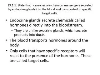
The pancreas is an organ in the digestive and endocrine system that serves two major functions: exocrine (producing pancreatic juice containing digestiveenzymes) and endocrine (producing several important hormones, including insulin). Pancreas
1: Head of pancreas2: Uncinate process of pancreas3: Pancreatic notch4: Body of pancreas5: Anterior surface of pancreas6: Inferior surface of pancreas7: Superior margin of pancreas8: Anterior margin of pancreas9: Inferior margin of pancreas10: Omental tuber11: Tail of pancreas12: Duodenum
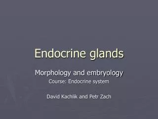
beta cells-Insulin and Amylin alpha cells-Glucagon Deltacells-Somatostatin PP cells-Pancreatic polypeptide 50-80%lower blood sugar 15-20%raise blood sugar 3-10%inhibit endocrine pancreas 1%inhibit exocrine pancreas There are four main types of cells in the islets of Langerhans.