Download
1 / 15
170 likes | 242 Vues
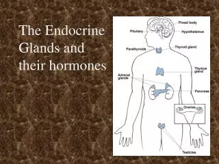
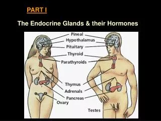
Explore the functions and anatomy of key endocrine glands like the ovaries, testes, adrenals, gut, kidneys, pancreas, thyroid, parathyroid, thymus, pituitary, hypothalamus, and pineal. Learn about hormones secreted and their roles in maintaining physiological balance. Dive into the intricate network of glands regulating various bodily functions and processes.

E N D
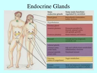
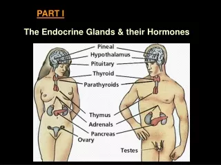
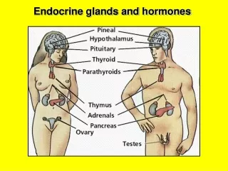
The ovaries The ovaries (A) are almond-shaped, bilateral glands about 4-6 cm long, located in the abdomen of the female. (B) A schematic diagram of a mammalian ovary showing several important anatomical features and the various stages of follicular and corpora luteal development. The insert shows different types of ovarian tissue as they are organized in the mature (Graafian) follicle, including the theca externa and interna, and granulosa cells. Prior to ovulation, the theca interna and follicular cells collaborate to synthesize estrogens. After ovulation, the cells comprising the follicle enlarge and differentiate into lutein cells which combine with theca interna cells to form the corpous luteum that secretes protestins. Hormones secreted: Progestins, androgens, estrogens, inhibin
The testes • A testis in cross section: • Showing seminiferous tubules • and vas deferens • (B) Seminiferous tubules in cross-section, • showing the heads of developing • spermatozoa embedded in Sertoli cells • And the steroid-making Leydig cells • In mammals, the testes begin as • undifferentiated gonads (like ovaries) • in the abdomen, but descend during • fetal development to be contained in • the scrotal sac, which is made out of • fused labial tissue. Hormones secreted: Progestins, androgens, inhibin
The adrenals The adrenal glands are found on top of each kidney. Each gland consists of two distinct regions, the cortex and medulla. The adrenal cortex also has cellular zones with different functional roles: The zona glomerulosa, marked by whorls of epithelial cells; the zona fasciculata, in which epithelial cells are organized in orderly bands; and the zona reticularis, where epithelial cells have a scattered appearance. Each gives rise to different secretions. Hormones secreted: Glucocorticoids (Cortisol) Mineralocorticoids (aldosterone) Progestins Adrenaline and Noradrenaline
The gut Consists of the pancreas, gall bladder, stomach, and duodenum. Involved in breakdown of food and extraction of water, electrolytes, minerals, and nutrients. MANY peptide hormones secreted, including insulin, glucagon, cholecystokinin (CCK), gastrin, and secretin, to regulate food breakdown and extraction, sugar and salt metabolism, electrolyte balance, thirst, feeding, and satiety. The kidneys Renin is released by granular cells of the juxtaglomerular apparatus in the medulla, which acts as an enzyme to create the peptide angiotensin (AII). In turn, AII acts on the subfornical organ in the brain to induce thirst, which restores blood volume and blood pressure.
The pancreas • (A) The pancreas is located beneath the liver • and rests in the curve of the duodenum, or • small intestine. • Most of the pancreas consists of exocrine • cells the secrete digestive fluids, islands of • endocrine tissue called Islets of Langerhans • are also present. • (C) Relative distribution of ɑ, β, and δ cells in • a pancreatic islet. Each type secretes a • different hormone. ɑ cells secrete glucagon β cells secrete insulin δ cells secrete somatostatin A fouth cell type, PP, secretes pancreatic polypeptide
The thyroid and parathyroid (~37C) The thyroid gland is a highly vascularized, H-shaped organ that partially surrounds the upper trachea. Embedded in each upper layer of the thyroid are two lobes of the parathyroid gland. The spherical, colloid-filled follicles of the thyroid synthesize, store, and secrete thyroid hormones.
The thymus The thymus is a specialized organ of the immune system that helps to create and sensitize T-cell lymphocytes through the action of thymosins, peptide hormones that stimulate lymphocyte production.
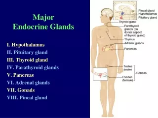
The hypothalamus (NOT A GLAND!) The hypothalamus is located at the base of the brain and consists of several collections of cell bodies called “nuclei”. Hypothalamic nuclei integrate hormonal, metabolic, and sensory information with descending control from limbic and cortical structures, and thereby coordinate various physiological processes. This occurs by means of special neurosecretory cells that project to the anterior or posterior pituitary.
The pineal The pineal gland is located between the telencelphaon and diencephalon. Its secretory cells are called pinealocytes, and secrete serotonin and melatonin. In fish, amphibians, reptiles, and birds, the pineal acts as an additional photoreceptor organ. In mammals, it is also responsive to light, and receives inputs from the retina. Melatonin regulates sleep-wake patterns, and seasonality.