Download
1 / 12
140 likes | 576 Vues
STUDY CASE # 1 - CYTOLOGY. INTERESTING LAB LLC 101 Beautiful St. Pomona, CA 88305 CYTOLOGY REPORT Patient Name: Vector, James DOS : 5/6/14 SPECIMEN 1: BLADDER, WASHING MICROSCOPIC DIAGNOSIS: Positive for malignancy.

E N D
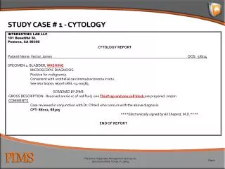
STUDY CASE # 1 - CYTOLOGY INTERESTING LAB LLC 101 Beautiful St. Pomona, CA 88305 CYTOLOGY REPORT Patient Name: Vector, James DOS: 5/6/14 SPECIMEN 1: BLADDER, WASHING MICROSCOPIC DIAGNOSIS: Positive for malignancy. Consistent with urothelial carcinoma/carcinoma in situ. See also biopsy report 1REL-14-00585. SCREENED BY DWR. GROSS DESCRIPTION: Received are 60 cc of red fluid; one ThinPrep and one cell block are prepared. zm/zm COMMENTS Case reviewed in conjunction with Dr. O’Neill who concurs with the above diagnosis. CPT: 88112, 88305 ****Electronically signed by Ali Sheperd, M.D.**** END OF REPORT
STUDY CASE # 2 – FINE NEEDLE ASPIRATION (FNA) • INTERESTING LAB LLC • 101 Beautiful St. • Pomona, CA 88305 • CYTOLOGY • Patient Name: Vector, James DOS: 5/6/14 • Final Diagnosis • Subcarina, fine needle aspiration biopsy: One thin preparation, one cell block, six Diff-Quik stained and four Papanicolaou stained smears are available for review. • Diagnostic category: Malignant. • Diagnosis: Poorly differentiated adenocarcinoma present. See comment. • Adequacy: Satisfactory for evaluation. • Comment • Immunohistochemical stains, performed with adequate controls on the cell block, show the carcinoma cells to be positive for CK7 and Napsin, focally positive for TTF-1 and CK5/6, and negative for p63 and CK20, supporting the diagnosis. • Page 1 of 2
STUDY CASE #2 – FINE NEEDLE ASPIRATION (FNA)(cont.) • CYTOLOGY NON-GYN REPORT • Patient Name: Vector, James DOS: 5/6/14 • Specimen(s) Received • 1 Subcarina • Clinical Information • Pre-op diagnosis: Lung mass • Post-op diagnosis: None given • Clinical History: Chest pain • Gross Description • Received are four alcohol-fixed smears for Papanicolaou stain and six Diff-Quik stained smears, and approximately 35 cc of pink, cloudy fluid fixed in CytoLyt which are submitted to Reliance. One ThinPrep and one cell block are additionally prepared and screened. • 5/5/2014 • Intraoperative Consultation • Preliminary impression regarding specimen adequacy per Dr. Sheperd. “Cells present suspicious for non-small cell carcinoma”. • CPT code: 88172, 88173, 88305, (88342 x 6 or G0461 and G0462x5) • ****Electronically signed by Ali Sheperd, M.D.**** • END OF REPORT
STUDY CASE #4 – FEMORAL HEAD PATHOLOGY REPORT Patient Name: Vector, James DOS: 5/6/14 SPECIMEN A, RIGHT FEMORAL HEAD COMPONENTS MICROSCOPIC DIAGNOSIS: “RIGHT FEMORAL HEAD”: FEMORAL HEAD WITH EBURNATION OF ARTICULAR CARTILAGE, AND FOCAL INTRAMEDULLARY HEMORRHAGE, CONSISTENT WITH FRACTURE. GROSS DESCRIPTION: The specimen is received in formalin labeled with the patient’s name and “right femoral head components” and consists of a fragmented, 5.0 x 4.5 x 4.0 cm femoral head which has a jagged, irregular, hemorrhagic bone resection margin. The cortical bone, articular surface is tan and smooth. Within the container is a 1.4 cm length of femoral neck which has one flat cut bone resection margin, and an opposing jagged, hemorrhagic irregular margin. The medullary bone is mottled from tan-red to tan-yellow and a tan articular rim ranges from 0.1 cm to 0.2 cm. A representative section from the jagged, hemorrhagic bone resection margin is submitted in one cassette, following decalcification. ****Electronically signed out**** Ali Shephard, M.D. CPT: 88305, 88311
STUDY CASE #5 - RENAL INTERESTING LAB LLC 101 Beautiful St. Pomona, CA 88305 SURGICAL REPORT Patient Name: Vector, James DOS: 5/6/14 Clinical History 37-year-old female with history of systemic lupus erythematosus is being evaluated for nephrotic range proteinuria and microscopic hematuria. Positive anti double stranded DNA, low C3 and C4. Creatinine 0.6. Urinalysis 10-20 WBCs, 30-50 red blood cells, ANA positive, 1:2560 homogeneous. Lupus anticoagulants negative. C3 21, C4 3.6, positive SSA. Positive Sm. Lupus diagnosis was made a few months ago and the patient has been treated with Prednisone and Plaquenil. Hematuria. Specimen(s) Received RIGHT KIDNEY BIOPSY Final Pathologic Diagnosis Needle biopsy of right kidney: Lupus nephritis. WHO Class III (B). ISN/RPS Classification: Class III-S (A/C). Minimal interstitial fibrosis and tubular atrophy. PHYSICIAN NOTIFICATION: The findings were discussed with Dr. Jones on 5/7/14. Dr. Smith was also notified at 4:30 p.m. on 4/10/14. ****Electronically signed by Ali Sheperd, M.D.**** page 1 of 3
STUDY CASE #5 – RENAL (cont.) • INTERESTING LAB LLC • 101 Beautiful St. • Pomona, CA 88305 • SURGICAL REPORT • Patient Name: Vector, James DOS: 5/6/14 • Gross Dictation • The specimen is retrieved fresh from CT by Dr. Picantie, labeled with the patient’s name and are three 0.1cm in diameter cores of tan tissue, 2.6 cm, 3.0 cm, and 3.1 cm. The specimen is submitted entirely as follows: • One 0.3cm in length core for electron microscopy, two cores (0.3 cm and 0.4 cm) for immunofluorescence studies and the remaining tissue (1.0 cm, 1.6 cm, 2.0 cm and 2.8 cm) for light microscopy in cassette KID. • INTRAOPERATIVE CONSULTATION: Adequate. Dr. Picantie • Intraoperative Consult Diagnosis • Adequate. Dr. Picantie • Microscopic Description • LIGHT MICROSCOPY (STAINS: H&E, PAS, PAMS, and MT): This biopsy is adequate and representative and consists of portions of cortex and medulla. At multiple levels, up to about 47 glomeruli are identified. Most of the glomeruli reveal mild mesangial expansion, predominantly with excess matrix and focally with slight increase in cellularity in some segments. In about 10 glomeruli, there is segmental architectural alterations with focal capillary luminal obliteration secondary to mesangial interposition and infiltration of leukocytes. There is minimal focal karyorrhexis but no evidence of fibrinoid necrosis. A rare segment shows adhesion to the capsule. Focal fibrosis is highlighted by Trichrome stain. • page 2 of 3
STUDY CASE #5 – RENAL (cont.) • INTERESTING LAB LLC • 101 Beautiful St. • Pomona, CA 88305 • SURGICAL REPORT • Patient Name: Vector, James DOS: 5/6/14 • Interstitium exhibits a few scattered foci of lymphocytic infiltration with associated focal mild interstitial fibrosis and tubular atrophy. The overall interstitial fibrosis and tubular atrophy is less than 2% of the examined cortex. The rest of the tubules are of normal size and morphology. The interlobular arteries are within normal limits. There is no microangiopathy or vasculitis. • DIRECT IMMUNOFLUORESCENCE: • H&E stained slides of the frozen section reveal a portion of renal cortex including up to about 8-10 glomeruli in different levels. 1+ granular peripheral capillary wall staining with focal mesangial staining is noted in for IgG (1+ in 4 glomeruli), C3 1+ in 6 glomeruli, Kappa (trace) in 5 glomeruli, Lambda 1+ in 6 glomeruli and C1q 1 to 2+ in 6 glomeruli. IgA, IgM, Fibrinogen and Albumin are negative. • ELECTRON MICROSCOPY: • Three blocks are prepared. Two glomeruli examined reveal multiple segments exhibiting small and medium-sized mesangial electron dense deposits and several subendothelial immune complex deposits. There are no epimembranous deposits. Mesangial interposition with duplication of basement membrane is identified focally. There are also scattered intraluminal leukocytes. A few foci of architectural disarray is noted with near total capillary luminal obliteration secondary to excess matrix, immune complex deposition and basement membrane disarray. Podocytes reveal a few foci of foot process simplification and effacement. • CPT: 88305, 88313 x 3, 88329 x 1, 88346 x 9, 88348 • END OF REPORT
STUDY CASE #6 – BONE MARROW • INTERESTING LAB LLC • 101 Beautiful St. • Pomona, CA 88305 • SURGICAL REPORT • Patient Name: Vector, James DOS: 5/6/14 • Clinical History • Bone depression to rule out bone marrow disorder. • Specimen(s) Received • BONE MARROW BIOPSY AND CLOT • Final Pathologic Diagnosis • THROMBOCYTOPENIA AND ANEMIA. • NORMOCELLULAR BONE MARROW WITH TRILINEAGE HEMATOPOIESIS. • ADEQUATE NUMBER OF MEGAKARYOCYTES. • Comment: The above findings are most consistent with the diagnosis of ITP. • Gross Description • The specimen is received in formalin labeled with the patient’s name and is a 1.0 cm in length x 0.2 cm in diameter core of tan to red-tan bone, with a 2.5 x 1.6 x 0.4 cm rectangular portion of red-brown clot. The specimen is submitted entirely as follows: • BM1) Bone core prior to decalcification. • BM2) Clot. • The procedure was performed by Dr. Ali Sheperd. • ****Electronically signed by Ali Sheperd, M.D.**** • page 1 of 3
STUDY CASE #6 – BONE MARROW • INTERESTING LAB LLC • 101 Beautiful St. • Pomona, CA 88347 • SURGICAL REPORT • Patient Name: Vector, James DOS: 5/6/14 • Microscopic Description • PERIPHERAL SMEAR • The red cells exhibit mild poikilocytosis with nucleated forms. The hemoglobin is 10.3 grams percent with an MCV of 77. The white count is 4700 with an automated differential of 55% polys, 39% lymphocytes and 6% monocytes. Atypical cells are not observed. The platelet count is 19,000. • BONE MARROW ASPIRATE: • The aspirate is cellular and adequate for evaluation. The differential consists of 2% blasts, 14% myelocytes, 15% metamyelocytes, 14% bands, 8% polyps, 21% lymphocytes and 26% normoblasts. Megakaryocytes are identified. Atypical cells are not observed. • BONE MARROW CLOT: • Marrow cellularity approximates 50%. Myeloid and erythroid cells are present in usual numbers. Moderate number of neutrophils are noted. Clusters of atypical lymphoid cells are not observed. Megakaryocytes are in their usual numbers with normal morphology. Granulomata and tumor cells are not present. An iron stain demonstrates absent iron stores. • BONE MARROW BIOPSY: • Bony trabeculae are unremarkable. The marrow cellularity approximates 50%. Blood vessels are normal. No evidence of granulomata or tumor. Megakaryocytes appear in usual numbers. • CPT: 38220, 38221, 85097, 88305 x 2, 88311, 88313
STUDY CASE #7 – GASTROINTESTINAL SPECIMEN INTERESTING LAB LLC 101 Beautiful St. Pomona, CA 88305 ANATOMIC PATHOLOGY REPORT Patient Name: Vector, James DOS: 5/6/14 SPECIMEN A, GASTRIC, BIOPSY MICROSCOPIC DIAGNOSIS: STOMACH, BIOPSY: PORTIONS OF SUPERFICIAL GASTRIC MUCOSA, ANTRAL AND FUNDIC TYPES, WITH MILD CHRONIC GASTRITIS. DIFF-QUIK STAIN FOR HELICOBACTER IS NEGATIVE. PLEASE SEE COMMENT. COMMENT: THE HISTOLOGIC FEATURES OF THIS GASTRITIS ARE SUGGESTIVE OF A CHEMCIAL TYPE GASTRITIS AS WOULD BE SEEN WITH THE USE OF NON-STEROIDAL ANTI-INFLAMMATORY DRUGS, ALCOHOL, OR WITH BILE REFLUX. CLINICAL CORRELATION IS SUGGESTED. GROSS DESCRIPTION: The specimen is received in formalin, labeled with the patient’s name and “gastric biopsy” and consists of two tan portions of soft tissue which are each 0.3 cm in greatest dimension. Both portions are submitted in toto in cassette A1. SPECIMEN B, ESOPHAGUS, BIOPSY MICROSCOPIC DIAGNOSIS:ESOPHAGUS, BIOPSY: PORTIONS OF SQUAMOUS MUCOSA WITH BASAL CELL HYPERPLASIA, SUGGESTIVE BUT NOT DIAGNOSTIC OF GASTROESOPHAGEAL REFLUX. AN ALCIAN BLUE/PAS STAIN DOES NOT REVEAL FUNAL ORGANISMS. GROSS DESCRIPTION: The specimen is received in formalin labeled with the patient’s name and “esophageal biopsy” and consists of two tan-gray portions of soft tissue which are 0.2 cm and 0.3 cm in greatest dimension. Both portions are submitted in toto in cassette B1. CPT: 88305 x 2, 88312 x 2 ****Electronically signed by Ali Sheperd, M.D.****


