Download
1 / 1
20 likes | 268 Vues
Olive oil polyphenols modify liver polar fatty acid composition and inhibit CCl 4 -induced hepatotoxicity in Balb/c mice. Jasminka Giacometti a , Hrvoje Križan a , Neven Franjić c and Alena Buretić-Tomljanović b

E N D
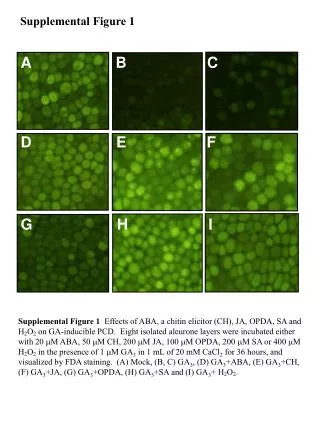
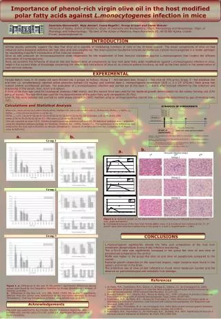
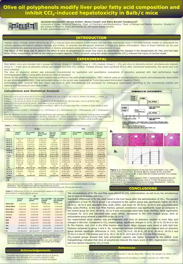
Olive oil polyphenols modify liver polar fatty acid composition and inhibit CCl4-induced hepatotoxicity in Balb/c mice Jasminka Giacomettia, Hrvoje Križana, Neven Franjićc and Alena Buretić-Tomljanovićb University of Rijeka, School of Medicine, aDept. of Chemistry and Biochemistry, bDept. of Biology and Medical Genetics, cStudent of the School of Medicine, Braće Branchetta 20, HR-51000 Rijeka, Croatia E-mail: jasminka@medri.hr INTRODUCTION Hepatic injury through carbon tetrachloride (CCl4) induced lipid peroxidation is well known and has been extensively used in the experimental models to understand the cellular mechanisms behind oxidative damage and further, to evaluate the therapeutic potential of drugs and dietary antioxidants. Many of these methods can be used often to study the potential preventive effect of dietary antioxidants when considering the measurements in vivo. The object of this study was to observe the effect of olive oil polyphenols on acute liver injury by determining the changes in the phospholipid (PL FAs) and free fatty acids (FFAs) composition, as well as the total antioxidant capacity (TEAC) of serum using the carbon tetrachloride (CCl4) induced liver injury on animal model. EXPERIMENTAL Male Balb/c mice were divided into 4 groups as follows: Group 1 - control; Group 2 - CCl4 treated; Group 3 - CCl4 and olive oil phenolics extract simultaneously treated; Group 4 – 3-day olive oil phenolics extract pre-treated and then CCl4 treated. Treated animals were sacrificed 24 hrs after mentioned treatments; the serum and liver were collected. The olive oil phenolics extract was previously characterized by qualitative and quantitative evaluations of phenolics analysed with high performance liquid chromatography (HPLC) using gallic acid as an internal standard. Serum PL FAs and FFAs fractions were isolated and purified by the solid-phase-extraction (SPE) method using an aminopropylsilica column and subsequently determined by gas chromatography (GC). Total antioxidant status in the serum was measured as Trolox Equivalent Antioxidant Capacity (TEAC). Immediately after removal, the liver tissue was fixed in 10% formaldehyde and processed for histological examination according to the conventional method and staining with hematoxylin and eosin (H&E). Calculations and Statistical Analysis SFAs=% (14:0+16:0+18:0+20:0+22:0+24:0); MUFAs=% (14:1+16:1+18:1+20:1);PUFAs=% (PUFAn-3 + PUFAn-6); PUFAs n–3 =% (20:5n-3+22:5n-3+22:6n-3); PUFAs n–6 =% (18:2n-6+18:3n-6+20:2n-6+20:3n-6+20:4n-6+22:4n-6);D9 C18 index =18:1n-9/18:03; D6D index=[(18:3n-6+20:3n-6)/18:2n-6]1; D5D index=[(20:4n-6/(20:3n-6)]1. ACL=[(% Total FAn x n)]/100 (n=carbon atom number)2; DBI= % of unsaturated FAs x number of duble bonds of each unsaturated FAs2; PI=[(%Monoenoic x 0.025) + (%Dienoic x 1) + (%Trienoic x 2) + (%Tetraenoic x 4) + (%Pentaenoic x 6) + (%Hexaenoic x 8)] 2; Data are reported as mean ±S.D. Statistical significance was assumed with p<0.05. All statistical analyses were conducted using the Statistica software package for Windows, StatSoft, Inc. (2005), Version 7.1. The significance of the differences was analyzed by the Descriptive Statistics by Groups (Breakdown) - Post-hoc Comparisons of Means using Scheffe test. Table 1. Differences in liver PL FAs and FFAs profiles between the control and test (treated) groups a. b. Figure 1.a. Histological analysis of the liver from male Balb/c mice. H&E-stained liver sections from: A-normal, B-CCl4 treated, C-CCl4 and OOP simultaneously treated and D- 3-days OOP pre-treated and then CCl4 treated mice, magnification = 400X; b.Serum total antioxidative capacity (TAC) * significantly different from the control and group 2, + from the group 2 and 3, # from the group 2 and 4, determined by the Descriptive Statistics by Groups (Breakdown) - Post-hoc Comparisons of Means using Scheffe test (p<0.05). Values are area per cent (mean±SD); *significantly different from the control determined by the Descriptive Statistics by Groups (Breakdown) - Post-hoc Comparisons of Means using Scheffe test (p<0.05). Abbreviations: PUFA-polyunsaturated fatty acid; MUFA-monounsaturated fatty acid; SFA-saturated fatty acid. CONCLUSIONS Table 2. Differences in liver PL characteristics based on FA profiles determined in control and test groups The concentrations of PL FAs and FFAs were altered by CCl4 administration, as well as by the simultaneous use of olive oil phenolics with CCl4. Significant differences in PL FAs were noted in the liver tissue after the administration of CCl4. The percent contribution of liver PL FAs in group 2 as compared to the control group was significantly higher for 18:0, 18:2n-6, 18:3n-6 and saturated fatty acids (SFA), and lower for 20:3n-6, 20:4n-6 and polyunsaturated fatty acids (PUFAs). In the liver FFAs fraction, percent contribution was significantly lower as compared to the control group for 14:0, 20:4n-6, 20:1n-9 and monounsaturated fatty acids (MUFAs), while significantly increased for 16:0 and saturated fatty acids (SFAs). Compared to the CCl4 treated group, olive oil antioxidants group showed a reduction in the 18:1n-9. Our experimental study showed that pre-treatment with olive oil phenolics resulted in more fatty acid changes in the liver. Compared to CCl4 treated group, significant changes in 14:0, 18:1n-9, 20:4n-6 in the PL FAs fraction, and 14:0 in the FFAs fraction were found. Major changes were noted in both lipid liver fractions compared to group 3 and 4. So, comparison between pre-treated and treated olive oil phenolics group showed significant differences in 14:0, 16:0, 18:1n-9, 18:3n-6, 20:3n-6, 20:4n-6, 20:5n-3 and 22:6n-6 in the PL FAs fraction and 14:0, 18:2, 20:3n-6, 24:0 and 22:6n-3 in the FFAs fraction. CCl4-treated groups showed that total antioxidant capacity was higher compared to the control. Liver histopathology indicated that olive oil polyphenols reduced the injury score of fatty degeneration incidence and liver lesions induced by CCl4 in mice. Values are area per cent (mean±SD); *significantly different from the control determined by the Descriptive Statistics by Groups (Breakdown) - Post-hoc Comparisons of Means using Scheffe test (p<0.05). Abbreviations: D9 C18-delta 9 desaturase index of the 18:0; D6D- delta 6 desaturase index; D5D-delta 5 desaturase index; ACL-average chain length; DBI-double bond index; PI-peroxidizability index. References Acknowledgements 1. Zuijdgeest-Van Leeuwen SD, Van der Heijden MS, Rietveld T, Van der Berg JWO, Tiltanus HW, Burgers JA, Wilson JHP, Dagnelie PC(2002) Br J Cancer 87: 1370 – 1378 2. Pamplona R, Portero-Otín M, Riba D, Ruiz C, Prat J, Bellmunt MJ and Barja G (1998) J Lipid Res 39: 1989–1994 This work was supported by theCroatian Ministry of Science, Project No. 062-0000000-0221 and 062-0982522-0369, DIOKI,d.d. Zagreb and The university of Rijeka foundation.