Download
1 / 29
320 likes | 1.09k Vues
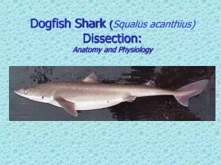
Dogfish Shark ( Squalus acanthius ) Dissection: Anatomy and Physiology. Dogfish Distribution. External Anatomy of the Dogfish Shark. Double dorsal fin anterior dorsal fin is larger than the posterior dorsal fin Presence two spines one immediately in front of each dorsal fin

E N D
Dogfish Shark (Squalusacanthius)Dissection:Anatomy and Physiology
External Anatomy of the Dogfish Shark • Double dorsal fin • anterior dorsal fin is larger than the • posterior dorsal fin • Presence two spines • one immediately in front of each dorsal fin • spines carry a poison secreted by glands at their base.
The paired pectoral fins • act like an airplane's wings to provide the lift needed to keep the shark from sinking. • The paired pelvic fins • located on either side of the cloacal aperture. They are different in males and females. • The cloacal opening • receives the products of the intestine, the urinary and the genital ducts. • The name cloaca, meaning sewer, seems quite appropriate.
Males have stout, grooved copulatory organs called claspers • During copulation, a clasperis inserted into the oviduct orifice of the female. The sperm proceed from the cloacaalong the groove on the dorsal surface of the clasper into the female. • The caudal finis divided into two lobes: a larger dorsal lobe and a smaller ventral lobe. This type of tail is known as a heterocercal tail.
eyes are prominent in sharks and are very similar to the eyes of man. • A transparent cornea covers and protects the eye. • Upper and lower eyelids protect the eye. • Just inside the lower lid is a nictating membrane that extends over the surface of the eye to cover the cornea.
Large spiracle openings are located posterior and dorsal to the eyes. • A valve, permits the opening and closing of the external spiracular pore. • The spiracle is an incurrent water passageway leading into the mouth for respiration.
Most sharks have five external gill slitslocated on the sides behind the mouth and in front of the pectoral fins. • Water taken in by the mouth and spiracles is passed over the internal gills and forced out by way of the gill slits.
The nares or external nostrils are located on the ventral surface of the rostrum anterior to the jaws. • Water passes into and out of the olfactory sac, permitting the shark to detect the odors of the water.
lateral line. • tiny pores on the side of the body that lead to receptors that are sensitive to the mechanical movement of water and sudden changes of pressure. • ampullae of Lorenzini • patches of pores on the head region • sensitive to changes in temperature, water pressure, electrical fields, and salinity.
Digestive Anatomy of the Dogfish Shark • A smooth, shiny membrane called peritoneumcan be seen lining the inside of the body wall. • The liver • largest organ • 3 lobes • two main lobes, the right and left lobes, extend from the length of the cavity. • A third lobe much shorter lobe contains the green gall bladder along its right edge.
Theesophagusis the thick muscular tube extending from the top of the cavity connecting the pharynx with the stomach. • The esophagus leads into the "J"-shaped stomach.
The duodenum is a short "U"-shaped portion of the small intestine that connects the stomach to the intestine. The bile duct from the gall bladder enters the duodenum. • The pancreas is located on the duodenum and the lower stomach. The secretions of the pancreas enter the duodenum by way of the pancreatic duct.
The dark, triangular-shaped spleen is located near the posterior end of the stomach. Although a part the circulatory system, the spleen is closely associated with the digestive organs in all vertebrates. • The valvular intestineis the second, and much larger, portion of the small intestine. It follows the duodenum and its outer surface is marked by rings.
The spiral valveis the screw-like, symmetrical shape within the valvular intestine. It adds surface area for digestion and absorption to an otherwise relatively short intestine.
The rectal glandis a slender, blind-ended, finger-like structure that leads into the intestine by means of a duct. It has been shown to excrete salt (NaCl) in concentrations higher than that of the shark's body fluids or sea water. It is thus an organ of osmoregulation, regulating the shark's salt balance. The cloaca is the last portion of the alimentary canal. It is a catch-all basin leading to the outside by means of the cloacal opening.
Respiratory Anatomy of the Dogfish Shark • The gills • composed of gill lamellae, blood vessels, and supporting cartilaginous structures located in a series of pharyngeal pouches. • provided with a rich blood supply. Arteries run directly from the nearby heart to the gills bringing deoxygenated blood into the gill lamellae. Oxygen diffuses from the ventilating water current flowing over the gills into the blood.
The pharynx is the portion of the alimentary canal posterior to the hyoid arch between the gills. Posteriorly it narrows to form the esophagus. • The spiraclesare openings in the anterior roof of the pharynx. The shark can bring water into its pharynx to the gills by way of the spiracle and mouth.
Urogenital Anatomy of the Dogfish Shark • Thekidneysare flattened, ribbon-like, darkly colored structures lying dorsally on either side of the midline, along the entire length of the body cavity.
Paired testes lie near the anterior end of the body cavity, dorsal to the liver, • The sperm pass from the testes to the kidneys within narrow tubules called efferent ductules. • Theovariesare two cream-colored elongated organs in the anterior part of the body cavity dorsal to the liver on either side of the mid-dorsal line. • The shape of the ovaries will vary depending upon the maturity of the specimen. • In mature specimens you may find two to three large eggs, about three centimeters in diameter, in each ovary.
Fertilizationis internal, usually taking place within the shell gland of the oviduct. The fertilized eggs continue to move posteriorly to the uterus. • As they grow the pups are attached to the egg, now known as the yolk sac. • During its period of gestation, which is nearly two years, the yolk is slowly absorbed by the shark "pup. • At birth the young are about 23 to 29 centimeters long. This type of development, where the young are born as miniature adults but have received hardly any nutrition directly from the mother's uterus, is known as ovoviviparous.