Dogfish Sharks
Dogfish Sharks. What phylum do Sharks belong to?. Characteristics of life of phylum chordata, class Chondrichthyes… genus squalus species acanthias. Cells: Multicellular Organization: Bilateral symmetry Require energy: predators, mainly boney fish… also canibalistic


Dogfish Sharks
E N D
Presentation Transcript
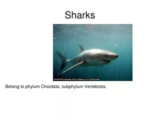
Dogfish Sharks What phylum do Sharks belong to?
Characteristics of life of phylum chordata, class Chondrichthyes… genus squalus species acanthias • Cells: Multicellular • Organization: Bilateral symmetry • Require energy: predators, mainly boney fish… also canibalistic • Responds: Complex nervous system, digestive system, etc.
Reproduction: Dioecious (males and females); mate in winter, ovoviviparous (eggs are incubated and hatched in females body) gestation is 22-24 months long. • Growth: Born at 20-30 cm long. Males reach maturity at age 11 and females at around 20 -35 years old. Females are larger than males and average 1 m in length at maturity.
Dogfish Shark (Squalus acanthius)Dissection:Anatomy and Physiology
External Anatomy of the Dogfish Shark • Along the sides of the body is a light-colored horizontal stripe called the lateral line. The line is made up of a series of tiny pores that lead to receptors that are sensitive to the mechanical movement of water and sudden changes of pressure. • The spiny dogfish has a double dorsal fin. The anterior dorsal fin is larger than the posterior dorsal fin. The spiny dogfish has the presence two spines, one immediately in front of each dorsal fin. The spines carry a poison secreted by glands at their base.
The caudal fin is divided into two lobes: a larger dorsal lobe and a smaller ventral lobe. This type of tail is known as a heterocercal tail. • The eyes are prominent in sharks and are very similar to the eyes of man. A transparent cornea covers and protects the eye. A darkly pigmented iris can be seen below the cornea with the pupil at its center. Upper and lower eyelids protect the eye. Just inside the lower lid is a membrane that extends over the surface of the eye to cover the cornea.
Large spiracle openings are located posterior and dorsal to the eyes. A spiracular valve, permits the opening and closing of the external spiracular pore. The spiracle is an incurrent water passageway leading into the mouth for respiration. • Most sharks have five external gill slits located on thire sides behind the mouth and in front of the pectoral fins. Water taken in by the mouth and spiracles is passed over the internal gills and forced out by way of the gill slits.
The paired pectoral fins act like an airplane's wings to provide the lift needed to keep the shark from sinking. • The paired pelvic fins are located on either side of the cloacal aperture. They are different in males and females.
The nares or external nostrils are located on the underside (ventral surface) of the rostrum anterior to the jaws. A nasal flap separates the incurrent from the excurrent opening. Water passes into and out of the olfactory sac, permitting the shark to detect the odors of the water. • The patches of pores on the head in the areas of the eyes, snout, and nostrils are the openings of the ampullae of Lorenzini. These sense organs are sensitive to changes in temperature, water pressure, electrical fields, and salinity.
Males have stout, grooved copulatory organs called claspers on the inner side of their pelvic fins. Fertilization in the dogfish shark is internal. During copulation, one of the claspers is inserted into the oviduct orifice of the female. The sperm proceed from the cloaca of the male along the groove on the dorsal surface of the clasper into the female.
The cloacal opening located on the ventral surface between the pelvic fins. It receives the products of the intestine, the urinary and the genital ducts. The name cloaca, meaning sewer, seems quite appropriate.
Digestive Anatomy of the Dogfish Shark • A smooth, shiny membrane called peritoneum can be seen lining the inside of the body wall. The visceral organs are suspended dorsally by a double membrane of peritoneum know as mesentery • The liver is the largest organ lying within the body cavity. Its two main lobes, the right and left lobes, extend from the pectoral girdle posteriorly most of the length of the cavity. A third lobe much shorter lobe is located medially and contains the green gall bladder along its right edge.
The esophagus is the thick muscular tube extending from the top of the cavity connecting the oral cavity and pharynx with the stomach. • The esophagus leads into the "J"-shaped stomach. The upper portion, the cardiac region, continues as the main body, and ends at the duodenal end.
The duodenum is a short "U"-shaped portion of the small intestine that connects the stomach to the intestine. The bile duct from the gall bladder enters the duodenum. • The pancreas is located on the duodenum and the lower stomach. The secretions of the pancreas enter the duodenum by way of the pancreatic duct.
The dark, triangular-shaped spleen is located near the posterior end of the stomach. Although a part the Iymphatic system, the spleen is closely associated with the digestive organs in all vertebrates. • The valvular intestine is the second, and much larger, portion of the small intestine. It follows the duodenum and its outer surface is marked by rings.
The spiral valve is the screw-like, symmetrical shape within the valvular intestine. It adds surface area for digestion and absorption to an otherwise relatively short intestine.
The colon is the narrowed continuation of the valvular intestine. It is located at the posterior end of the body cavity. • The rectal gland is a slender, blind-ended, finger-like structure that leads into the colon by means of a duct. It has been shown to excrete salt (NaCI) in concentrations higher than that of the shark's body fluids or sea water. It is thus an organ of osmoregulation, regulating the shark's salt balance.
The cloaca is the last portion of the alimentary canal. It collects the products of the colon as well as the urogenital ducts. It is a catch-all basin leading to the outside by means of the cloacal opening.
Respiratory Anatomy of the Dogfish Shark • The gills are the respiratory organs of the shark. They are composed of gill lamellae, blood vessels, and supporting cartilaginous structures are are located in a series of pharyngeal pouches. • The oral cavity is the area enclosed by the jaws (mandibular arch) and the cartilage of the throat (hyoid arch). • The tongue of the shark is practically immovable and without muscles. It is supported anteriorly and posteriorly by cartilage.
The pharynx is the portion of the alimentary canal posterior to the hyoid arch between the gills. Posteriorly it narrows to form the esophagus. • The spiracles are openings in the anterior roof of the pharynx. The shark can bring water into its pharynx to the gills by way of the spiracle and mouth.
The gills are provided with a rich blood supply. Arteries run directly from the nearby heart to the gills bringing deoxygenated blood into the gill lamellae. Oxygen diffuses from the ventilating water current flowing over the gills into the blood
Cardiovascular • Closed circulatory system • 2 chambered heart (atrium & ventricle) • Sinus venous collects blood from the veins • Blood then is pumped to atrium, then ventricle • Then blood is pumped to conus arteriousus • Blood then is pumped to gills to be oxygenated, from gills to the rest of the body • Deoxygenated blood is drained from the body by veins and returned to the caridnal sinuses, then the cardinal veins, then the sinus venosus
Urogenital Anatomy of the Dogfish Shark • The urinary and genital systems have distinct and unique functions. The first, the removal of nitrogenous wastes and the maintenance of water balance; the other, the reproduction of species. However, due to their similar developmental origins and the sharing of common structures, they are usually considered as a single system. • The shark kidney and its ducts are quite different from those in higher vertebrates. The relationship between the urinary and genital structures is also quite different.
The kidneys are flattened, ribbon-like, darkly colored structures Iying dorsally on either side of the midline, along the entire length of the body cavity. A tough white glistening strip of connective tissue is found between the kidneys in the midline. • The kidneys of the male are essentially the same as those of the female. The posterior portion is involved in the manufacture and transport of urine. The main difference lies in the anterior portion of the kidney, which in females is degenerate and functionless, but in males is an active part of the reproductive system.
Paired testes lie near the anterior end of the body cavity, dorsal to the liver, adjacent to the anterior ends of the kidneys.The sperm pass from the testes to the kidneys within narrow tubules called efferent ductules. • After passing through the anterior end of the kidney the sperm enter the ductus deferens and pass posteriorly toward the cloaca. In mature male specimens the ductus deferens may be seen on the ventral surface of the kidneys as a pair of highly coiled tubules. • Note: While in the female this duct carries urine, in the male it transports spermatozoa and seminal fluid.
The claspers are modified extensions of the medial portions of the pelvic fins. They are inserted into the female's cloaca during copulation. • The finger-like claspers each have a dorsal groove, the clasper tube that carries the seminal fluid from the cloaca of the male to the cloaca of the female during mating.
The ovaries are two cream-colored elongated organs in the anterior part of the body cavity dorsal to the liver on either side of the mid-dorsal line. The shape of the ovaries will vary depending upon the maturity of the specimen. In immature females they will be undifferentiated and glandular in appearance. In mature specimens you may find two to three large eggs, about three centimeters in diameter, in each ovary. When these break the surface of the ovary, upon ovulation, they enter the body cavity and by means of peritoneal cilia are moved into the oviducts.
Fertilization in the dogfish shark is internal, usually taking place within the shell gland of the oviduct. The fertilized eggs continue to move posteriorly to the uterus. As they grow the pups are attached to the egg, now known as the yolk sac, by means of a stalk. During its period of gestation, which is nearly two years, the yolk is slowly absorbed by the shark "pup."Numerous uterine villi, finger-like projections from the uterine wall, make contact with the surface ot the developing embryo and its yolk sac. It is believed that these provide the embryo with water; all other nutrients are supplied by the yolk. At birth the young are about 23 to 29 centimeters long. This type of development, where the young are born as miniature adults but have received hardly any nutrition directly from the mother's uterus, is known as ovoviviparous.