Understanding FNAB and Imaging Techniques for Breast Cancer Diagnosis
100 likes | 225 Vues
This document outlines the role of Fine Needle Aspiration Biopsy (FNAB) in evaluating breast mass lesions, highlighting its procedure using a fine gauge needle to sample fluid or tissue. It describes the diagnostic significance of mammography for early breast cancer detection, chest X-rays for monitoring metastasis, and ultrasound for differentiating cysts and solid masses. Additionally, it touches on CT scans and bone scans in assessing cancer spread and conditions affecting bones. The document concludes with insights on proceeding to core biopsy if FNAB results are negative.

Understanding FNAB and Imaging Techniques for Breast Cancer Diagnosis
E N D
Presentation Transcript
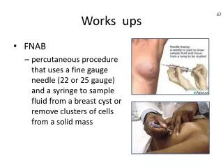
Works ups • FNAB • percutaneousprocedure that uses a fine gauge needle (22 or 25 gauge) and a syringe to sample fluid from a breast cyst or remove clusters of cells from a solid mass
FNAB This type of biopsy is performed for one of two reasons: - A biopsy is performed on a lump or a tissue-mass when its nature is in question. - For known tumors, this biopsy is performed to assess the effect of treatment or to obtain tissue for special studies.
Mammography • process of using low-dose amplitude-X-rays to examine the human breast and is used as a diagnostic and a screening tool. • early detection of breast cancer, typically through detection of characteristic masses and/or microcalcifications. Screening: 40y/o every 1-2years 50y/o once a year
Mammography Malignant lesions tend to have irregular and (usually) spiculated margins Normal Fatty breast Benign well-defined edges and a halo sign
IN OUR PATIENT Irregular and spiculatedmargins
Chest X-ray • to check and see whether the cancer has metastasize to the lungs During treatment for breast cancer: • If a person has advanced breast cancer that has spread to the lungs, a chest x-ray is used to check on how the disease is responding to treatment. • For people who develop a fever during chemotherapy, chest x-rays are used to check for the presence of pneumonia. • If a person experiences new shortness of breath in the first few months after radiation therapy, with or without a cough, to see if the radiation caused any inflammation of the lungs.
Ultrasound • method of choice for the differentiation of cysts from solid masses and for guidance in interventional procedures. • Benign: • solid lesions with smooth or lobulated margins that are sharply defined, with homogeneous hypoechoic contents and a horizontal orientation • Malignant: • hypoechoic lesions with irregular and poorly defined margins the lesion is a fibroadenoma
CT scan • may be used as an adjuvant for monitoring spread • effective for the detection of intraductal extension of breast carcinoma and is thought to be useful in the preoperative assessment of indications of breast-conserving surgery.
Bone scan • nuclear scanning test that identifies new areas of bone growth or breakdown • It can be done to evaluate damage to the bones, find cancer that has metastasizedto the bones • monitor conditions that can affect the bones BONE SCAN FINDINGS Confirmed osteoblastic metastatic bone lesions that enhance on radionuclide bone scan
If FNAB is negative, what will you do? • CORE BIOPSY • procedure where a needle is passed through the skin to take a sample of tissue from a mass or lump. • more invasive procedure than FNAB, as it involves a local anaesthetic.