TKA in The Valgus Knee
1.2k likes | 4k Vues
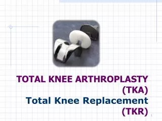
TKA in The Valgus Knee. Andrew Tice Arthroplasty Rounds March 8, 2012 Credit: Greg Hansen 2008. TKA for Valgus Deformity. Much less common than varus deformity Approximately 10-15% of all TKA more common in: Females Rheumatoid Arthritis Previous trauma Renal osteodystrophy

TKA in The Valgus Knee
E N D
Presentation Transcript
TKA in The Valgus Knee Andrew Tice Arthroplasty Rounds March 8, 2012 Credit: Greg Hansen 2008
TKA for Valgus Deformity • Much less common than varus deformity • Approximately 10-15% of all TKA • more common in: • Females • Rheumatoid Arthritis • Previous trauma • Renal osteodystrophy • Rickets • Poliomyelitis
TKA for Valgus deformity • The deformity is defined as > 5-7 degrees of tibio femoral valgusangulation • Comprised of: • Bone loss from lateral femoral condyle • Contractures of lateral soft-tissue structures • Attenuated MCL • Instability
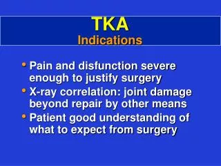
Indications • Patients with: • End-stage degenerative joint disease • Pain • Limitation of activities of daily living • Increasing angular deformity • Increasing instability • Other non-operative (mechanical or pharmacological) therapies have failed
Contraindications • Absolute • Infection • Relative • Young age • High activity level • Obesity
Physical Exam • Analyze gait • Establish presence of deformity (varus/valgus, flexion contracture) • Evaluate ROM • Evaluate Stability • Evaluate Patellofemoral tracking • Evaluate Extensor Mechanism Function • Document Neurovascular examination Radiographic Examination • Standard Weight bearing AP,Lateral, full length alignment, and Skyline Patellar Views
Surgical Goals • Remain the same as with Varus Deformity • Pain Relief • Restore alignment • Restore stable joint • Provide functional ROM • Provide proper patellofemoral tracking • Stable well-fixed prosthesis
Valgus-Specific Complication • Peroneal Nerve Palsy • Rates of 3-4 % • Most commonly due to traction from overcorrection of valgus deformity with a thick spacer • May also occur when performing posterior capsule release by pie-crusting • Nerve may be relaxed with flexion of the knee
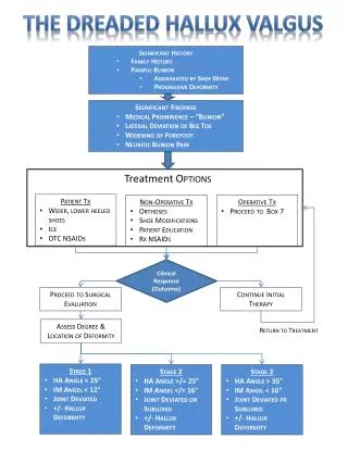
Classification – Krackow et al. • Variant 1 • 80% of cases • <10 degrees • Passively correctable • Intact MCL
Classification – Krackow et al. • Variant 2 • 15% • 10-20 degrees • Not correctable with varus stress • MCL elongated but functional
Classification – Krackow et al. • Variant 3 • 5% • >20 degrees • Not corretable with varus stress • MCL attenuated • Marked lateral soft tissue contracture
Surgical Approach • Medial Parapatellar approach remains the most common approach • Lateral Parapatellar approach is associated with concerns regarding soft tissue coverage following release • Previously found to have comparable results in achieving correction of alignment
Surgical Approach • 44 patients (22 Med, 22 Lat + TTO) • No sig. difference in IKSS score, IKSS functional score, ROM, VAS score, number of knees with valgus deformity • Did sig. differ with regards to degree of deformity (IKSS scoring system) • Lat + TTO: 2 knees 9-10 degrees from ideal range • Med: 7 knees 10-13 degrees from ideal range
Bone Resection • Distal Femoral cut at 4-5 degrees instead of 5-7 degrees for varusknee (less dev. from mechanical axis – full length films) • Tibial Resection made at 90 degree angle to axis of shaft of tibia and with posterior slope • Anterior/Posterior cuts more difficult due to hypoplastic lateral condyle • with posterior condylar reference guide, will be pushed into internal rotation • Based off Whiteside’s line, transepicondylar line (3 degrees of ER WRT femoral shaft) • Judged with posterior condylar axis and tibial shaft axis • Checked by symmetric flexion gap with soft tissues corrected
Soft Tissue BalancingLateral Release • Many different methods/sequences proposed • 3 lateral Layers • Superficial: deep fascia – IT band, biceps tendon, lateral gastrocs • Middle: Lateral Retinaculum, patellofemoral ligament • Deep: Capsule, Arcuate Ligament, LCL and Popliteus Tendon
Soft Tissue BalancingLateral Release • First remove all osteophytes • Extension space addressed initially: • Release of ITB: at Gerdy’s tubercle, lengthening with multiple stab incisions (pie-crusting), or at joint line • If more correction needed release posterior capsule: pie-crusting • Awareness of peroneal nerve 7-9 mm from capsule • Gastrocs and Biceps release a rare option
Soft Tissue BalancingLateral Release • Flexion gap addressed next • Release LCL: anterior to posterior • Posterior lateral corner • Popliteus tendon: resect or secure to LCL to avoid joint block from hanging tendon
Soft Tissue BalancingMedial Release • If Medial ligamentous stretching or attenuation, may need to address medial side as well • Advancement of MCL • Division and imbrication
Other Considerations • PCL may need to be sacrificed with elevating the joint line, secondary to soft tissue release, or stretched due to valgus deformity • Posterior stabilized component • Posterior stabilized, constrained component • Only true consideration in elderly, low function
Other Considerations • Patellofemoral tracking • Lateral release • TibialTuberosity transfer
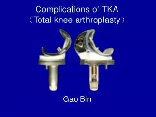
Complications • Soft-tissue instability with risk of dislocation • 6-24% • Patellar maltracking • 0-4% • Peroneal nerve Palsy • 0-4% • Wound Complications • 1-10% • Recurrent Valgus Deformity • 5-20%
References • Lombardi, AV et al. An algorithmic approach to TKA in the valgus knee. JBJS 2004, 86:62-71 • Williot, A et al. Total Knee Arthroplasty in the Valgus Knee. Orthopedics and Traumatology2010, 96S: S37-S42 • Peters, CL et al. Primary Total Knee Arthroplasty in the Valgus Knee. The Journal of Arthroplasty 2001, 16: 723-729 • Meek, RM et al. Lateral Ligament Release to Correct Valgus Deformity During Surgery. Techniques to Knee Surgery 2003: 2(1): 18-27 • Healy, WL et al. Medial Reconstruction During Total Knee Arthroplasty for Severe Valgus Deformity 1998, 356: 161-169 • Nikolopoulos DD et al. Total Knee Arthroplasty in severe valgus knee deformity: comparison of a standard medial parapatellar approach combined with tibial tubercle osteotomy . Knee Surg Sports TraumatolArthrosc 2011: 19: 1835-1842