Download
1 / 2
20 likes | 198 Vues
3D GaN-Ga 2 O 3 Core Shell Structures Revealed by X-ray Diffraction Microscopy Jianwei Miao, UCLA, DMR 0520894.

E N D
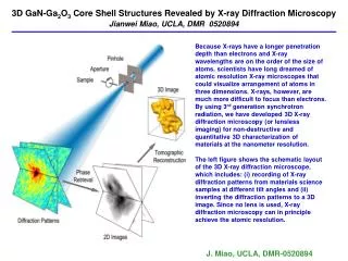
3D GaN-Ga2O3 Core Shell Structures Revealed by X-ray Diffraction Microscopy Jianwei Miao, UCLA, DMR 0520894 Because X-rays have a longer penetration depth than electrons and X-ray wavelengths are on the order of the size of atoms, scientists have long dreamed of atomic resolution X-ray microscopes that could visualize arrangement of atoms in three dimensions. X-rays, however, are much more difficult to focus than electrons. By using 3rd generation synchrotron radiation, we have developed 3D X-ray diffraction microscopy (or lensless imaging) for non-destructive and quantitative 3D characterization of materials at the nanometer resolution. The left figure shows the schematic layout of the 3D X-ray diffraction microscope, which includes: (i) recording of X-ray diffraction patterns from materials science samples at different tilt angles and (ii) inverting the diffraction patterns to a 3D image. Since no lens is used, X-ray diffraction microscopy can in principle achieve the atomic resolution. J. Miao, UCLA, DMR-0520894
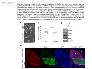
3D GaN-Ga2O3 Core Shell Structures Revealed by X-ray Diffraction Microscopy Jianwei Miao, UCLA, DMR 0520894 By using the X-ray diffraction microscope, we, for the first time, carried out quantitative 3D imaging of a heat-treated GaN particle with each voxel corresponding to 17 17 17 nm3. We observed the platelet structure of GaN and the formation of small islands on the surface of the platelets, and successfully captured the internal GaN-Ga2O3 core shell structure in three dimensions. This work opens the door for non-destructive and quantitative imaging of 3D morphology and 3D internal structure of a wide range of materials at the nanometer scale resolution that are amorphous or possess only short-range atomic organization. The up-left figures show the iso-surface renderings of a reconstructed 3D GaN quantum dot particle, (A) the front view, (B) the back and (C) the side view. (D) shows 3D internal structure of the GaN quantum dot particle with each slice of 17 nm. The 3D GaN-Ga2O3 core shell structure is visible where the low electron density corresponds to -Ga2O3 and the high density corresponds to the GaN cores. Miao et al., “Three-Dimensional GaN-Ga2O3 Core Shell Structures Revealed by X-ray Diffraction Microscopy ”, Phys. Rev. Lett., 97, 215503 (2006). Featured in National Cancer Institute, United Press International, the Materials World Magazine, Medical News Today A B C D