Download
1 / 31
320 likes | 775 Vues
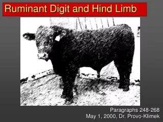
Ruminant Digit and Hind Limb. Paragraphs 248-268 May 1, 2000, Dr. Provo-Klimek. http://www.bleatingheart.com/jacobsheep.html. http://www.jacobsheep.com/. Review.

E N D
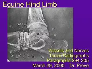
Ruminant Digit and Hind Limb Paragraphs 248-268 May 1, 2000, Dr. Provo-Klimek
http://www.bleatingheart.com/jacobsheep.html http://www.jacobsheep.com/
Review • You should review the dissection of the proximal part of the hind limb (PP 195-213). We will not cover this in lecture today, but you are responsible for all previous dissection of the hind limb. • You are also responsible for all previous information regarding the hind limb skeleton.
Review - innervation • Make sure you know the areas of skin supplied by the following: • Dorsal branches of lumbar and sacral nerves • Cutaneous branches of the pudendal nerve/caudal cutaneous femoral nerve • Lateral cutaneous femoral nerve • Saphenous nerve • Tibial and common peroneal nerves • Note that tibial and common peroneal together innervate all the structures of the limb distal to the stifle (except for skin innervated by saphenous).
Foot, Digits, Claws and Dewclaws foot (Dyce) digits or toes dewclaw (hoof only) fetlock jt. pastern jt. coffin jt. bulb (heel) sole wall claws (hoof)
Example Quiz Questions • Identify this part of the hoof. • Bulb or heel • What is the difference between the bovine and equine laminae of the hoof/dermis? (Golden Oldie from Micro…) • Ruminants only have primary laminae. • Identify this junction between the sole and wall. • White line IV III IV
Common and Lateral Digital Extensor Tendons common digital extensor: Note: “just like” the horse, but double because 2 digits. medial head lateral digital extensor lateral head Note: three palpable extensor tendons, rather than two as in the horse. Dorsal view: IV III L M
Ligaments of Digits distal (deep, cruciate) sesamoidean ligs. interdigital phalangeo-sesamoidean ligs. proximal interdigital lig. distal interdigital lig. (deep and superficial parts; superficial part crosses DDF tendon and thus acts like an annular ligament) Note the absence of middle and superficial distal sesamoidean ligaments. (More on that later.)
Superficial and Deep Digital Flexor, Ligaments (See ND 744 for similar diagram) DDF SDF SDF DDF interdigital ligs: proximal distal palmar annular lig. digital annular ligs. Lateral view Palmar view
Interosseous Medius Attachments - Palmar View Note: “just like” the horse, but double because 2 digits. extensor tendon attachment to prox. sesamoids extensor branch (abaxial) extensor branch (axial) dist. sesamoid. lig. palmar lateral
Slip of Interosseous to SDF Tendon deep digital flexor superficial digital flexor interosseous slip attachment to proximal sesamoids abaxial extensor branch The attachment of the interosseous to the SDF functions in place of the superficial distal sesamoidean ligament. lateral
Interosseous Medius Attachments - Dorsal View Note: “just like” the horse, but double because 2 digits. lateral head of common digital extensor lateral digital extensor medial head of common digital extensor (blue) extensor branches of interosseous: abaxial axial L M
Example Quiz Questions • How many extensor tendons are palpable: • here? • Three • here? • Three
Ligaments and Tendons of Digits interosseous slip to SDF tendon axial extensor branch prox. interdigital lig. annular ligaments dist. interdig. lig.
Hind Limb Arteries - Summary • Dorsal metatarsal artery III is largest supply to foot. • continuation of cranial tibial/dorsal pedal • perforating arteries detached to plantar surface in proximal and distal metatarsus • joins with plantar common digital artery III, and together these supply the axial digital arteries • Saphenous artery divides into small medial and lateral plantar arteries. • these divide into plantar common digital arteries • plantar common digital artery III is largest and vulnerable to laceration at fetlock
Hind Limb Arteries saphenous dorsal pedal dorsal MT III plantar common digital III (47)
Break: Example Quiz Questions • If this giraffe fractured its metatarsal bone at the level indicated: • what major artery might be severed? • branches of the nerve on either side of the flexor tendons would be vulnerable to injury. • the interosseous (would/would not) be vulnerable here. Dorsal MT III tibial
Hind Limb Nerves - Summary • Tibial and common peroneal nerves supply limb distal to tarsus. • Common peroneal: • deep branch supplies dorsal metatarsal nerve • superficial branch supplies dorsal common digital nn. II, III and IV • Tibial nerve: • divides into medial and lateral plantar, which further divide into plantar common digital nn. II, III and IV • Digital nerves in hind limb follow same pattern as those in forelimb.
Hind Limb Nerves tibial medial plantar lateral plantar deep br. peroneal sup. branches peroneal plantar dorsal
Four-point block in mid-metatarsus same puncture • on both sides of flexor tendons between the tendons and interosseous: • medial and lateral plantar nerves • on dorsal side between MT bone and extensor tendons: • deep peroneal nerve (dorsal metatarsal nerve) • on dorsal side on either side of the cranial branch of lateral saphenous vein: • superficial peroneal nerve branches 4 1 2 3 plantar dorsal
Landmarks • Lateral saphenous • cranial branch goes to the dorsal surface of the metatarsus • Interosseous • Digital flexor tendons • (palpate)
Injection Sites for Digital Anesthesia Note: this will not block the fetlock or the metatarsus because of cutaneous branches given off higher in the metatarsus. Lateral plantar medial plantar Superficial and deep peroneal ( deep peroneal)
Mid-Metatarsal Nerve Block deep peroneal n. medial plantar n. LoDE DDF IM SDF LaDE superficial peroneal n. lateral plantar n.
Nerve Block at Metacarpo(tarso)phalangeal Joint • Dorsal and palmar abaxial digital nerves on the medial and lateral digits: • inject 1/2 - 1 cm. dorsal to dewclaw on medial and lateral side of foot • Dorsal axial digital nerves: • inject on the dorsal midline just distal to the metacarpo(tarso)phalangeal joint • Palmar axial digital nerves: • inject on the palmar (plantar) midline at the level of the dewclaws
Muscle Miscellany • Bovine reciprocal apparatus. • it’s there - review it from the horse discussion • Peroneus tertius is muscular. • most cranial muscle of crus • covers both cranial tibial and long digital extensor • Long digital extensor arranged similarly to common digital extensor. • medial belly goes to digit 3 (P2 and P3) • lateral belly goes to digit 3 and 4 (P3)
Spastic Paresis • Periodic spastic contraction of the gastrocnemius. Hock extended (also stifle to some extent – reciprocal apparatus). Typically only when standing. • Etiology unknown. Heritability controversial. • Signs usually in cattle younger than 1 year. • Treatment by surgical interruption of the tibial nerve fibers to the gastrocnemius.
Example Quiz Questions • Name a muscle responsible for extension of this joint. • Quadriceps • Gluteobiceps • Long digital extensor /peroneus longus • Identify the nerve that supplies this area of skin. • Common peroneal