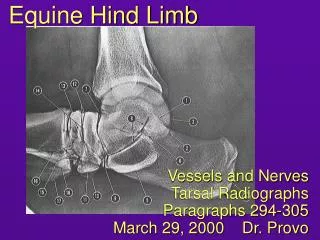
Equine Hind Limb
Equine Hind Limb. Reciprocal Apparatus, Stifle, Tarsus and Hip Paragraphs 306-313 March 30, 2000. Announcements. Do paragraphs 306-311 on one leg (the best specimen), and do paragraphs 312-313 on the other. There will be a short “one-minute paper” at the end of the lecture today.

Equine Hind Limb
E N D
Presentation Transcript
Equine Hind Limb Reciprocal Apparatus, Stifle, Tarsus and Hip Paragraphs 306-313 March 30, 2000
Announcements • Do paragraphs 306-311 on one leg (the best specimen), and do paragraphs 312-313 on the other. • There will be a short “one-minute paper” at the end of the lecture today.
Reciprocal Apparatus • Involves fibrous bands which unite the distal femur to the tarsus/metatarsus. • peroneus tertius on the cranial side • superficial digital flexor and fibrous band in the gastrocnemius on the caudal side • Functions to cause the stifle and hock to move in unison. • when the stifle flexes, the hock flexes; when the stifle extends, the hock extends • Bottom line: if the stifle is fixed in position, the hock is fixed also.
Peroneus Tertius and Cranial Tibial S447 • Peroneus tertius (1 - yellow) • dorsal insertion on T3 and MT III • lateral insertion on calcaneus and T4 • Cranial tibial (2 - blue) • perforates distal end of the peroneus tertius • cunean tendon inserts medially on T 1&2 • cunean bursa • dorsal insertion on MT III M ND622 M
Caudal Leg Muscles 1. gastrocnemius 2. popliteus soleus 3. deep digital flexor 4. lateral digital extensor 5. superficial digital flexor location of calcaneal bursa ND623 S449
Reciprocal Apparatus patella femur tibia superficial digital flexor peroneus tertius calcaneus talus metatarsus From the Mediclip image bank
Reciprocal Apparatus standing
Reciprocal Apparatus flexed
Comprehension Break: Examples (Extreme) flexion of both stifle and hock. Flexion of stifle with extension of hock.
Example Quiz Questions 1. The calcanean tendon keeps the hock from (flexing, extending) while the horse is bearing weight on that limb. 2. What part of the reciprocal apparatus is located here (cranial side of crus)? Peroneus tertius
Stay Apparatus of the Hind Limb • Same principle as that for the forelimb: • horse is able to stand with minimal muscular effort required to keep limb joints from collapsing • Key components: • patella and patellar ligaments (locks stifle) • caudal structures of the crus/tarsus (stabilize tarsus when stifle is locked) • tarsal ligaments, attachment of SDF to calcaneus, DDF with its check ligament, common calcanean tendon (gastrocnemius) • suspensory apparatus of the fetlock (same as forelimb) • Bottom line: fixation of the stifle allows fixation of the tarsus; digit has suspensory apparatus.
Patellar Ligaments and Locking Mechanism Patella in resting pos. medial fibrocartilage medial lig. (1’) middle lig. (1’’) lateral lig. (1’’’) Patella slides on gliding surface during movement. Patellar fibrocartilage and medial patellar ligament “lock” over medial trochlear ridge. This is an active process - involves extension, & medial rotation of patella S365
Stifle Joint - Medial View (ND 616) 1. patella 2. patellar fibrocartilage 4. med. trochlear ridge 5. middle patellar lig. 6. medial patellar lig. These structures form the locking mechanism of the stifle; “2” and “6” loop over the medial trochlear ridge. medial
Stifle Joint - Lateral and Caudal (S368-9) med. patellar ligament biceps femoris femoropatellar lig. cran. cruciate lig. lat. collateral meniscofemoral lat. meniscus caud. cruciate lig. lat. patellar lig. lateral
Successive stages of patellar movement: 1 2 3 3 1 2
Ligaments of the Tarsus (S371-2) 1. collateral ligs. 1”. short parts of 1 2. long plantar lig. lateral medial area of cunean bursa dorsal tarsal lig
Passive Stay Apparatus • Hip is relatively balanced. • Stifle locked with patellar ligaments. • Weight of trunk would tend to flex hock. • flexion prevented by tension in superficial digital flexor (red) and gastrocnemius (blue) • peroneus tertius (red) not involved in standing animal • stay apparatus of digit essentially same as that of forelimb • notice check lig. for deep digital flexor (green) force of weight
Stifle Joints (S366) • Stifle consists of the femoropatellar joint and the femorotibial joint. • femorotibial joint has both medial and lateral compartments • femoropatellar usually communicates with the medial femorotibial compartment; occasionally it also communicates with the lateral • in practice, treat all stifle joint compartments separately
Tib. F Cal. Talus central 4 1+2 3 II III IV Tarsal Bones and Joints (after Dyce) Note: schematic diagram share a joint capsule tarsocrural proximal row proximal intertarsal intermediate row distal intertarsal distal row tarsometatarsal occasionally communicate
Tarsocrural Joint Pouches dorsomedial lateroplantar medioplantar M deep dig. flexor lateral malleolus medial malleolus (and ‘3’) (easily punctured, but avoid saphenous v.)
Synovial Structures of Tarsus (ND 620) L cross section at arrows in A medial
Synovial Structures of Tarsus - Key 1. superficial digital flexor and associated bursae 2. gastrocnemius 3. deep digital flexor (and tarsal sheath in tarsal canal) 5. medial saphenous vein (cr. branch) 6. long digital extensor 7. peroneus tertius 8. cranial tibial (cunean tendon - 8’) 9. proximal, middle and distal extensor retinacula 10. tarsocrural joint with two of three pouches showing 11. collateral ligaments 14. long plantar ligament 15. plantar nerves and saphenous vessels 16. cranial tibial vessels and deep peroneal nerve 18. caud. cut. sural nerve, lat. saphenous vein
Ligaments of Hip - Cranial View (S441) transverse acetabular ligament prepubic tendon accessory ligament of head of femur (orange) (round ligament of head of femur is not shown)
Example Quiz Questions 1. (Breed?) 2. Which patellar ligaments can be palpated? 3. What artery is palpable here? Holsteiner All 3 can be palpated. (Lateral plantar) digital
One-Minute Paper (complete and turn in) 1. What topic was presented the most clearly today? 2. What topic was least well presented? 3. What would have improved today’s lecture?
1. What topic was presented the most clearly today? 55 Reciprocal apparatus / animated pictures 12 Patellar locking mechanism / stay apparatus 4 Everything / well done 1 Ligaments of hip 2. What topic was least well presented? 31 Synovial structures of tarsus / stifle 13 Stay apparatus / patellar locking or movement 8 Nothing / nothing listed 7 Accessory ligament of head of femur / hip ligaments 3 Tarsal ligaments 2 Blood flow (yesterday) 1 Topics later in the lecture What would have improved today’s lecture? 29 Nothing, nothing listed or well done 10 More time for the hind limb labs / add one more lab / spread out material 8 More time on synovial structures 5 More time to draw on notes, especially radiographs / slow down 5 Better handout (although “better than most”); list / label more structures / color copies 3 Schematic or model of locking mechanism / review stay apparatus 2 Fewer tangents / less repetition in lecture 2 More real pictures, less illustrations 2 Sites for hock injections / hock diseases Miscellaneous: beer, more sleep (2), a Quarter Horse; leave ½ of the room lights on; shorter lectures; table to differentiate components of common calcanean tendon; don’t ask quiz questions from next assignment; orient to illustrations before discussing them One-Minute Paper Results, 3-29-00