Download
1 / 8
80 likes | 100 Vues
Learn the anatomy of a long bone and how to interpret X-rays for diagnosing bone injuries. Discover why doctors need multiple X-rays for accurate diagnoses. Participate in a gallery walk to showcase your mastery of X-ray analysis.

E N D
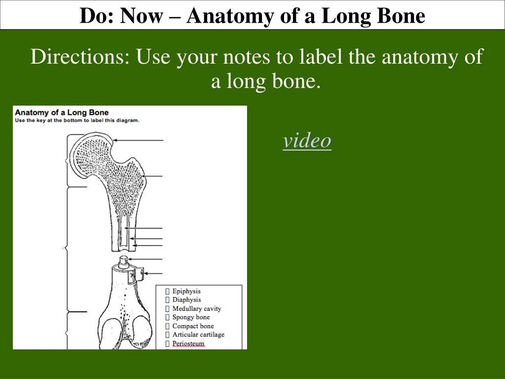
Do: Now – Anatomy of a Long Bone Directions: Use your notes to label the anatomy of a long bone. A video
Lab: Identifying X-Rays Objective: Diagnose bone injuries based on x-ray analysis. Provide 2 pieces of evidence to support the claim “Doctors need more than one x-ray to correctly diagnose bone injuries.” READ and ANNOTATE. Answer the Checking for Understanding Questions #1 – 2
Lab: Identifying X-Rays Objective: Diagnose bone injuries based on x-ray analysis. Provide 2 pieces of evidence to support the claim “Doctors need more than one x-ray to correctly diagnose bone injuries.” READ and ANNOTATE. Answer the Checking for Understanding Questions #3
Lab: Identifying X-Rays Objective: Diagnose bone injuries based on x-ray analysis. Provide 2 pieces of evidence to support the claim “Doctors need more than one x-ray to correctly diagnose bone injuries.” READ and ANNOTATE. Answer the Checking for Understanding Questions # 4 - 6
Each group will create and present a SMALL POSTER based on analysis of 2 patient X-Rays. • All groups will participate in Gallery Walk presentations to demonstrate mastery of SWBAT • Diagnose bone injuries based on x-ray analysis. Lab: Identifying X-Rays – gallery walk
Include the following on your poster: • Pictures of x-rays • (DO NOT REMOVE FROM PLASTIC) • Both normal and abnormal images • Answers to 5 questions (from your lab) • appendicular/axial or both • Names of bones • Type of injury/fracture • Abnormal findings • Cause of injury Lab: Identifying X-Rays – gallery walk
Gallery Walk Expectations • Everyone presents (2 rounds) • Everyone participates • Complete questions for patients • (4 patients are optional) • Answer Keys will provided after the gallery walk • Team Leaders – See Ms. Fine before your group presents! Lab: Identifying X-Rays – gallery walk
Provide evidence to support the claim. • See the x-rays of patients with more than 1 x-ray. • Patient 1 • Patient 6 • Patient 9 • Patient 11 Lab: Identifying X-Rays – Written Assessment


