Lung Cancer
Lung Cancer. Overview. Mortality trends Risk factors Screening Solitary pulmonary nodule Staging /Survival by stage Histology Molecular Testing Presentations/patterns of spread Paraneoplastic phenomena Management of resectable disease Adjuvant therapy Neoadjuvant therapy

Lung Cancer
E N D
Presentation Transcript
Overview • Mortality trends • Risk factors • Screening • Solitary pulmonary nodule • Staging /Survival by stage • Histology • Molecular Testing • Presentations/patterns of spread • Paraneoplastic phenomena • Management of resectable disease • Adjuvant therapy • Neoadjuvant therapy • Management of unresectable disease
Lung cancer mortality trends Lung cancer “epidemic” - has peaked in men – appears to have leveled in women Peter Jennings Vincent Schiavelli Don Knotts Dana Reeves Joe Paterno
2011 Estimated US Cancer Cases* Men822,300 Women774,370 Prostate 29% Lung & bronchus 14% Colon & rectum 9% Urinary bladder 6% Melanoma of skin 5% Non-Hodgkin lymphoma 4% Kidney & renal pelvis 5% Leukemia 3% Oral cavity 3% Pancreas 3% All Other Sites 19% 30% Breast 14% Lung & bronchus 9% Colon & rectum 6% Uterine corpus 4% Non-Hodgkin lymphoma 4% Melanoma of skin 5% Thyroid 3% Kidney & renal pelvis 3% Ovary 3% Pancreas 22% All Other Sites *Excludes basal and squamous cell skin cancers and in situ carcinomas except urinary bladder. Source: American Cancer Society, 2011.
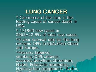
LUNG CANCER Epidemiology • Estimated 239,320 in U.S. 20111 • 128,890 male • 110,430 female • Most common cancer overall • 2nd most common cancer in men and women • Leading cause of cancer related death (161,250 deaths) • Also leading cause of cancer related death worldwide
Lung cancer – risk factors • Cigarette smoking accounts for about 90% of all lung cancer • Increased use increases risk: 40 pk-yr = 20xRR of a non-smoker • Corollary: reduction from 2PPD to ½ PPD will reduce risk • Environmental (second-hand) tobacco smoke increases risk • Radiation therapy • RT after breast ca – increased Lung Cancer among smokers – ipsilateral lung • RT for Hodgkin’s Lymphoma assoc w/increased risk of Lung Cancer • Caveat: Improved RT techniques to reduce exposure of lung is hoped to reduce this complication in the future
Lung cancer – risk factors • Other known/ascribed risk factors • Radon (emanation from soil - unpredictable) • Asbestos • Metals (arsenic, chromium, nickel) • Ionizing radiation (occupational/accidental) • Pulmonary fibrosis (independent of smoking) • Polycyclic aromatic hydrocarbons (inhaled from incomplete combustion - auto pollution, cooking oils, soot) • HIV infection • Genetic factors – Family History = 2x risk (after controlling for smoking) • Major susceptibility locus (chromosome 6q23–25) for inherited lung cancer
Screening – National Lung Screening Trial (NLST) • Diagnosis of lung cancer generally based upon evaluation of individuals with symptoms. • Screening for lung cancer has not been widely used • CXR and sputum cytology not shown to reduce lung ca mortality • National Lung Screening Trial (NLST) • Multicenter (33 medical centers) • Screening of high-risk pts for 3 yrs, N=53,454 • Age 55-74, “High-risk” = 30+ pack-years (allowed prior use if quit within 15 yrs of enrollment) • Annual Low-dose chest CT versus CXR • (+) findings = noncalcified nodule ≥4 mm on CT scan or any noncalcified nodule on x-ray.
Screening – National Lung Screening Trial (NLST) • Interim analysis 11/2010 benefit for CT scanning at a Median follow-up of 6.5 years CT group CXR group • (+) screen 24% 6.9% • False (+)/complication rate 96.4% / 1.4% 94.5% / 1.6% • Cases/100K person 645 572 • Stage I/II at dx 70% 56.7% • Lung Ca deaths 247 309 (Relative Risk Reduction 20%) • Conclusions: NLST demonstrated that CT screening reduced mortality in a high-risk population, compared to screening by x-ray • Number needed to screen w/CT to prevent one lung cancer death was 320 • Cost per life saved high, given high false-positive rate and subsequent w/u • NELSON trial is a randomized CT-based lung cancer trial being conducted in the Netherlands and Belgium; CT screening is being compared to no screening in 7,557 current or former smokers
Tumor is the RumorIs Cancer the Answer? • Solitary pulmonary nodule represents potentially curable stage of lung cancer • Stage I Lung cancers are within the definition of a SPN • Goal – identify and resect potentially curable cancer and avoid surgical resection of benign nodules • The SPN also represents a host of other “non-malignant” processes • CT scan with fine cuts through nodule helpful to characterize Size Low Risk High Risk Per Up-To-Date Jan2012 • 4-6 mm 12 12, 24 Repeat scan (months) • 6 to 8 mm 12, 24 6, 12, 24 • >8 mm 3, 9, 24 3, 9, 24 • A nodule that has clearly grown on serial imaging tests should be excised • If > 1 cm … FDG-PET sensitivity of 95% / specificity 78% for malignancy
Heartworm! Tissue is the ISSUE! ?
NSCLC TNM definitions - 2011 Tumor Characteristics • T1: 3cm, surrounded by lung/visceral pleura, not in main bronchus T1a 2, T1b >2-3 cm • T2: >3 to 7 cm, or tumor involving: • Main bronchus involvement and 2 cm distal to the carina • Visceral pleura invasion • Assoc w/ atelectasis or obstructive pneumonitis extending to hilar region but not involve the entire lung T2a > 3 but 5, T2b > 5 to 7 • T3: Tumor > 7 cm or involves • Chest wall (including superior sulcus), diaphragm, phrenic nerve, mediastinal pleura, or parietal pericardium • Main bronchus and < 2 cm distal to the carina but without involvement of the carina • Assoc atelectasis or obstruct pneumonitis of entire lung • Separate/multiple nodules in same lobe • T4: Tumor invading: • Mediastinum, heart, great vessels, trachea, esophagus, vertebral body, carina; • Separate tumor nodules in different ipsilateral lobe TOO MUCH TO COMMIT TO MEMORY – LOOK THIS UP IN NCCN GUIDELINES
NSCLC TMN (Continued) Regional lymph nodes (N) • N0: No regional lymph node metastasis • N1: Ipsilateral peribronchial, intrapulmonary, hilar • N2: Ipsilateral LN within the mediastinal and/or subcarinal • N3: Contralat mediastinal, contralat hilar, any scalene, or SC LN(s) Distant metastasis (M) • M0: No distant metastasis • M1: Distant metastasis • M1a Separate tumor nodule(s) in contralateral lobe or malignant effusion (pleural or pericardial) • M1b Distant mets TOO MUCH TO COMMIT TO MEMORY – LOOK THIS UP IN NCCN GUIDELINES
Staging NSCLC – 7th Edition TOO MUCH TO COMMIT TO MEMORY – LOOK THIS UP IN NCCN GUIDELINES
Non Small Cell Lung Cancer Most people are diagnosed Stage III and IV. 25% stage I 7% stage II 32% stage III 36% stage IV 70% with Stage I-III, will have their disease recur
Histologic Classification Keratinization and intercellular bridges c/w SCC • Squamous cell carcinoma (20%) • Decreasing incidence - ? Filters - smaller particles or having to suck moves smoke to the periphery • Adenocarcinoma (38%) • bronchioloalveolar • F>M and non-smokers • acinar , papillary , solid with mucus formation • Small cell carcinoma (13%) • Other variants (29%) • Large cell carcinoma (5%) • Spindle cell variant • Giant cell • Clear cell • Undifferentiated carcinoma • Other NOS Adenocarcinoma Bronchioloalveolar – well-differentiated columnar cells proliferating along the framework of alveolar septae. Small cell - cells are almost only blue nucleus (DNA) material making them "small" under the microscope
Genetics of NSCLC Hecht S S JNCI J Natl Cancer Inst 1999;91:1194-1210 • Smoking causes many chromosomal abnormalities • Del 3p – ~90% Small cell Ca and ~50% NSCLC • Del 8p (21.3-22) ~ 50% NSCLC • Deletions and point mutations p53 gene • (loss of inhibition of proliferation) • Loss of PTEN (inhibits PI3K/Akt pathway) • - PI3Ks-Akt constitutively activated in NSCLCs • K Ras mutations • highly assoc w/ resistance to TKIs • lack of response to platinum/vinorelbine treatment
Required Molecular TestingEpidermal Growth Factor Receptor (EGFR) • Del Exon 19 or point mutation in Exon 21 confer EGFR TKI sensitivity • Exon 20 mutations associated with resistance to EGFR TKI • More common Asians, females, non-smokers – REMEMBER IT! • EGFR TKIs appropriate to use front line if mutation present Mok TS, et al. N Engl J Med 2009;361: 941-57. Janne, ASCO Educational Session, 2007 Pooled results from 7 trials
Required Molecular TestingEML4-ALK translocations • Identified in small subset of NSCLC (5-7%) – does not overlap with kRas or EGFR • Fusion protein – Can be detected FISH, RT-PCR, or IHC • Vast majority tend to be adenocarcinoma (some reports in SqCC) • Tend to be younger, non-smokers/light smokers • Associated with response to Crizotinib Kwak et al. N Engl J Med 2010;363:1693-703.
Lung Cancer Mutation Consortium: Incidence of single-driver mutations NO MUTATIONDETECTED KRAS22% AKT1 NRAS MEK1 MET AMP EGFR17% HER2 PIK3CA BRAF 2% Mutation found in 54% (280/516) of tumors completely tested (CI 50-59%) DOUBLE MUTANTS 3% EML4-ALK7% Kris MG et al. Proc ASCO 2011;Abstract CRA7506.
Occult presentations Asymptomatic pulmonary nodule Lung mass/mediastinaladenopathy Subacute/Insidious Presentations Unexplained weight loss Non-resolving /post-obstructive PNA New onset clubbing New hoarseness (recurrent laryngeal nerve) DRAMATIC PRESENTATIONS SVC syndrome (flushing/ headache/plethora) Pericardial/Pleural effusion New onset Hypercalcemia Seizure/weakness from CNS met Pain C8, T1, T2 distro (Pancoasttumor) Tumor expectoration (uncommon but exciting!) Panoply of Presentations – seen them all ...
Panoply of Presentations – seen them all ... • Common/Typical Presentations • Cough (New or Worsening chronic cough) (50-75%) • Hemoptysis (25-50%) • Chest pain from local invasion (20%) • Increased DOE (obstruction with collapse/effusion) (25%) • Bone pain – back > rib > pelvis
Squamous Centrally located, can be associated with necrosis Bronchoalveolar Patchy infiltrates -Spreads along airways Adenocarcinoma Often more peripheral lesions Small cell Mediastinal adenopathy Early and WIDELY metastatic disease Tissue Is the Issue but . . .“Typical” Patterns of disease
Bone Liver Brain Adrenal Patterns of metastasis
Hypercalcemia • Most common in SQUAMOUS CELL CA • Polyuria, metab alkalosis, hyperuricemia, ARF • Hypomotility with anorexia, N/V, constipation, also pancreatitis • Weakness/lethargy/coma/seizures. • Can suggest bony mets or PTH-related-peptide • PTHrP binds PTH receptors Ca2+mobilization • TX: Volume repletion, saline diuresis, bisphosphonate, effective treatment of the tumor
Hypertrophic Osteoarthropathy • Primarily seen in NSCLC – rare in SCLC • DOES NOT INDICATE METASTASIS • HPO - subperiostealcancellous bone at distal ends of long bones. • Radius and ulna (80%) or tibia/fibula (74%). • Sx’s: pain, swelling, erythema • Long bone x-rays can show subperiosteal bone formation • Clubbing • Bone scan activity long bones • Etiology ? Neurogenic / humoral. • Neurogenic theory – vagotomy can result in ipsilateral remission. • Humoral theory - ? substance related to the malignancy - resection of tumor can result in immediate symptom relief.
Paraneoplastic syndromesSmall Cell Lung Cancer • Cushing's syndrome due to excess ACTH • SIADH • Lambert-Eaton myasthenic syndrome • Cerebellar ataxia, subacute sensory neuropathy • THESE ARE ASSOC WITH SMALL CELL CANCER • TOMORROW’S LECTURE WITH DR. CARTER!
Decision making • Resectability – surgery remains the foundation of therapy • Resectable surgery +/- adjuvant • “Potentially” resectable due to primary “neoadjuvant” • Convert necessary surgery from a pneumonectomy (which the patient cannot tolerate) to a lobectomy (which the patient could tolerate) • “Potentially” resectable due to specific mets • Solitary mets to adrenal • Mets to different lobes in unilateral lung • Solitary brain mets – resection of primary as well as metastatectomy with additional whole-brain XRT – prolonged survival • Unresectable • Poor PS due to comorbidities or inadequate lung function • Bulky mediastinal (N2) adenopathy • Contralateral disease • Metastatic to bilateral lung, bone, liver, or brain (with exceptions above)
Stage I-IIIA Can the patient tolerate surgery? • Rule of thumb – PFT’s can suggest what surgery a patient can tolerate • Pneumonectomy – FEV1 2.0 L • Lobectomy – FEV1 1.5 L • Better to use post-operative % predicted FEV1 • Postop FEV1 at least 800 cc and FEV1 predicted >40% • Split function testing (nuc-med) helps estimate postop function • Marginal pts: get CPEX: if VO2 max > 15 will tolerate most resections w/ normal M&M • Limited resection – segmentectomy (preferred) or wedge resection can be considered • Low DLCO predicts increased morbidity and need for post-resection home O2 • Mortality is different based on location!! • Right Pneumonectomy 12% Mortality • Left Pneumonectomy 6% Mortality
Survival by stage s/p curative resection Van Rens M et al Chest:2000:117:374 Betticher DC, Lung Ca: 2005: S9-16
Post-surgery treatment (Adjuvant) • Improved disease free survival • Improved overall survival absolute JBR-10, NEJM, 352;25, 2005 IALT, NEJM 350;4, 2004 ANITA, LancetOnc, 2006
Post-surgery treatment (Adjuvant) • Meta-analysis of studies • Disease free survival HR 0.84 (0.78-0.91) • Overall Survival HS 0.89 (0.82-0.96) • Absolute overall survival benefit at 5 yrs = 5.4% J Clin Oncol. 2008 Jul 20;26(21):3552-9.
Post-surgery treatment (Adjuvant) • For Stage IA disease – generally observation • Caveat: positive margins – re-resection or RT • For Stage IB-III disease – Adjuvant chemotherapy • “Platinum based” doublet • Cisplatin (preferred) or carboplatin • Combined with another agent • vinorelbine, etoposide, vinblastine, gemcitabine, paclitaxel, docetaxel, or pemetrexed • May seem random, but decision is based off of comorbidities, histology, patient preference, physician preference • If positive margins – chemo-RT followed by chemo
What about patients who have large tumors with local invasion (i.e.; surgery, if possible, is going to leave tumor at the margins)? Superior sulcus Chest Wall Invasion Mediastinal Invasion
Management of local invasion • If it appears to be resectable • Preoperative concurrent chemotherapy with radiation followed by surgery . . . Then adjuvant chemo
Pre-op Chemo-RTPhase III RTOG 9309 (INT 0139) • 396 patients, PS 0-1, Stage IIIA (T1–3N1-3M0) • All received concurrent chemoRT (45Gy) • Cisplatin 50mg/m2 D1, 8 x 2 cycles q28 d • Etoposide 50 mg/m2 D1-5 x 2 cycles q28 d • Randomization: Surgery vs Definitive RT up to 61 Gy • All received 2 more cycles of Cis-Etoposide • Early treatment related mortality • Surgery 7.9% vs RT 2.1% Lancet. 2009 Aug 1;374(9687):379-86.
Pre-op Chemo-RTPhase III RTOG 9309 (INT 0139) Surgery RT • Median OS 23.6 mo 22.2 mo (p=NS) • 5-yr PFS 21% 11% (p=0.008) • 5-yr OS 27% 20% (p=NS) • Pneumonectomy 26% postop mortality • Lobectomy 1% postop mortality Conclusions: surgery after chemo-RT can be considered in fit pts requiring lobectomy Concurrent Chemo-Radiation should continue uninterrupted if pneumonectomy would be required Lancet. 2009 Aug 1;374(9687):379-86.
Management of UNRESECTABLE disease • Chemotherapy with radiation • Concurrent treatment > sequential • About 20-25% OS at 5 years Curran WJ, J Natl Cancer Inst. 2011 Oct 5;103(19):1452-60. Lancet. 2009 Aug 1;374(9687):379-86.
Management of metastatic disease Schiller JH. N Engl J Med. 2002 Jan 10;346(2):92-8. • Complete discussion beyond the scope of this talk • IM Board testable questions • “Platinum based doublet” is the standard front line regimen • EXCEPT EGFR sensitive mutation – should receive EGFR Tyrosine Kinase inhibitor (e.g., erlotinib) • EXCEPT EML-ALK translocation – may receive crizotinib frontline • EXCEPT elderly or extensive comorbidities – single agent regimens • No benefit of systemic chemotherapy for ECOG PS 3-4
Management of metastatic disease • Important concepts – but probably not board testable • Bevacizumab marginal improvement added to doublet – but only for NON-SQUAMOUS • Cetuximab marginal improvement added to doublet – regardless of histology • Platinum-pemetrexed may be more efficacious for adenoca • Platinum-gemcitabine may be more efficacious for squamous cell ca • Maintenance therapy after initial chemo delays progression and improves OS (but is it just early second line therapy?)
Management of metastatic disease • Second line therapy and beyond – all dependent on performance status • Single agent therapy • Pemetrexed • Docetaxel • Paclitaxel or Nab-paclitacel • Erlotinib • Vinorelbine