Download
1 / 84
890 likes | 1.18k Vues
21. P A R T A. The Immune System: Innate and Adaptive Body Defenses. Immunity: Two Intrinsic Defense Systems. Innate (nonspecific) system responds quickly and consists of: First line of defense – skin and mucosae prevent entry of microorganisms

E N D
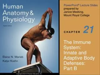
21 P A R T A The Immune System: Innate and Adaptive Body Defenses
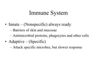
Immunity: Two Intrinsic Defense Systems • Innate (nonspecific) system responds quickly and consists of: • First line of defense – skin and mucosae prevent entry of microorganisms • Second line of defense – antimicrobial proteins, phagocytes, and other cells • Inhibit spread of invaders throughout the body • Inflammation is its most important mechanism
Immunity: Two Intrinsic Defense Systems • Adaptive (specific) defense system • Third line of defense – mounts attack against particular foreign substances • Takes longer to react than the innate system • Works in conjunction with the innate system
Innate and Adaptive Defenses Figure 21.1
Surface Barriers • Skin, mucous membranes, and their secretions make up the first line of defense • Keratin in the skin: • Presents a physical barrier to most microorganisms • Is resistant to weak acids and bases, bacterial enzymes, and toxins • Mucosae provide similar mechanical barriers
Epithelial Chemical Barriers • Epithelial membranes produce protective chemicals that destroy microorganisms • Skin acidity (pH of 3 to 5) inhibits bacterial growth • Sebum contains chemicals toxic to bacteria • Stomach mucosae secrete concentrated HCl and protein-digesting enzymes • Saliva and lacrimal fluid contain lysozyme • Mucus traps microorganisms that enter the digestive and respiratory systems
Respiratory Tract Mucosae • Mucus-coated hairs in the nose trap inhaled particles • Mucosa of the upper respiratory tract is ciliated • Cilia sweep dust- and bacteria-laden mucus away from lower respiratory passages
Internal Defenses: Cells and Chemicals • The body uses nonspecific cellular and chemical devices to protect itself • Phagocytes and natural killer (NK) cells • Antimicrobial proteins in blood and tissue fluid • Inflammatory response enlists macrophages, mast cells, WBCs, and chemicals • Harmful substances are identified by surface carbohydrates unique to infectious organisms
Phagocytes • Macrophages are the chief phagocytic cells • Free macrophages wander throughout a region in search of cellular debris • Kupffer cells (liver) and microglia (brain) are fixed macrophages Figure 21.2a
Phagocytes • Neutrophils become phagocytic when encountering infectious material • Eosinophils are weakly phagocytic against parasitic worms • Mast cells bind and ingest a wide range of bacteria
Mechanism of Phagocytosis • Microbes adhere to the phagocyte • Pseudopods engulf the particle (antigen) into a phagosome • Phagosomes fuse with a lysosome to form a phagolysosome • Invaders in the phagolysosome are digested by proteolytic enzymes • Indigestible and residual material is removed by exocytosis
Microbe adheres to phagocyte. 1 Phagocyte forms pseudopods that eventually engulf the particle. 2 Phagocytic vesicle containing antigen (phagosome). Lysosome Phagocytic vesicle is fused with a lysosome. 3 Phagolysosome Microbe in fused vesicle is killed and digested by lysosomal enzymes within the phagolysosome, leaving a residual body. 4 Acid hydrolase enzymes Residual body Indigestible and residual material is removed by exocytosis. 5 (b) Figure 21.2b
Microbe adheres to phagocyte. 1 (b) Figure 21.2b
Microbe adheres to phagocyte. 1 Phagocyte forms pseudopods that eventually engulf the particle. 2 (b) Figure 21.2b
Microbe adheres to phagocyte. 1 Phagocyte forms pseudopods that eventually engulf the particle. 2 Phagocytic vesicle containing antigen (phagosome). Lysosome (b) Figure 21.2b
Microbe adheres to phagocyte. 1 Phagocyte forms pseudopods that eventually engulf the particle. 2 Phagocytic vesicle containing antigen (phagosome). Lysosome Phagocytic vesicle is fused with a lysosome. 3 Phagolysosome Acid hydrolase enzymes (b) Figure 21.2b
Microbe adheres to phagocyte. 1 Phagocyte forms pseudopods that eventually engulf the particle. 2 Phagocytic vesicle containing antigen (phagosome). Lysosome Phagocytic vesicle is fused with a lysosome. 3 Phagolysosome Microbe in fused vesicle is killed and digested by lysosomal enzymes within the phagolysosome, leaving a residual body. 4 Acid hydrolase enzymes Residual body (b) Figure 21.2b
Microbe adheres to phagocyte. 1 Phagocyte forms pseudopods that eventually engulf the particle. 2 Phagocytic vesicle containing antigen (phagosome). Lysosome Phagocytic vesicle is fused with a lysosome. 3 Phagolysosome Microbe in fused vesicle is killed and digested by lysosomal enzymes within the phagolysosome, leaving a residual body. 4 Acid hydrolase enzymes Residual body Indigestible and residual material is removed by exocytosis. 5 (b) Figure 21.2b
Natural Killer (NK) Cells • Can lyse and kill cancer cells and virus-infected cells • Are a small, distinct group of large granular lymphocytes • React nonspecifically and eliminate cancerous and virus-infected cells • Kill their target cells by releasing perforins and other cytolytic chemicals • Secrete potent chemicals that enhance the inflammatory response
Inflammation: Tissue Response to Injury • The inflammatory response is triggered whenever body tissues are injured • Prevents the spread of damaging agents to nearby tissues • Disposes of cell debris and pathogens • Sets the stage for repair processes • The four cardinal signs of acute inflammation are redness, heat, swelling, and pain
Inflammation Response • Begins with a flood of inflammatory chemicals released into the extracellular fluid • Inflammatory mediators: • Kinins, prostaglandins (PGs), complement, and cytokines • Released by injured tissue, phagocytes, lymphocytes, and mast cells • Cause local small blood vessels to dilate, resulting in hyperemia
Toll-like Receptors (TLRs) • Macrophages and cells lining the gastrointestinal and respiratory tracts bear TLRs • TLRs recognize specific classes of infecting microbes • Activated TLRs trigger the release of cytokines that promote inflammation
Inflammatory Response: Vascular Permeability • Chemicals liberated by the inflammatory response increase the permeability of local capillaries • Exudate—fluid containing proteins, clotting factors, and antibodies • Exudate seeps into tissue spaces causing local edema (swelling), which contributes to the sensation of pain
Inflammatory Response: Edema • The surge of protein-rich fluids into tissue spaces (edema): • Helps dilute harmful substances • Brings in large quantities of oxygen and nutrients needed for repair • Allows entry of clotting proteins, which prevents the spread of bacteria
Inflammatory Response: Phagocytic Mobilization • Four main phases: • Leukocytosis – neutrophils are released from the bone marrow in response to leukocytosis-inducing factors released by injured cells • Margination – neutrophils cling to the walls of capillaries in the injured area • Diapedesis – neutrophils squeeze through capillary walls and begin phagocytosis • Chemotaxis – inflammatory chemicals attract neutrophils to the injury site
Innate defenses Internal defenses Positive chemotaxis 4 Inflammatory chemicals diffusing from the inflamed site act as chemotactic agents Diapedesis Neutrophils enter blood from bone marrow 3 1 Margination 2 Endothelium Basement membrane Capillary wall Figure 21.4
Innate defenses Internal defenses Inflammatory chemicals diffusing from the inflamed site act as chemotactic agents Neutrophils enter blood from bone marrow 1 Figure 21.4
Innate defenses Internal defenses Inflammatory chemicals diffusing from the inflamed site act as chemotactic agents Neutrophils enter blood from bone marrow 1 Margination 2 Endothelium Basement membrane Capillary wall Figure 21.4
Innate defenses Internal defenses Inflammatory chemicals diffusing from the inflamed site act as chemotactic agents Diapedesis Neutrophils enter blood from bone marrow 3 1 Margination 2 Endothelium Basement membrane Capillary wall Figure 21.4
Innate defenses Internal defenses Positive chemotaxis 4 Inflammatory chemicals diffusing from the inflamed site act as chemotactic agents Diapedesis Neutrophils enter blood from bone marrow 3 1 Margination 2 Endothelium Basement membrane Capillary wall Figure 21.4
Antimicrobial Proteins • Enhance the innate defenses by: • Attacking microorganisms directly • Hindering microorganisms’ ability to reproduce • The most important antimicrobial proteins are: • Interferon • Complement proteins
Interferon (IFN) • Genes that synthesize IFN are activated when a host cell is invaded by a virus • Interferon molecules leave the infected cell and enter neighboring cells • Interferon stimulates the neighboring cells to activate genes for PKR (an antiviral protein) • PKR nonspecifically blocks viral reproduction in the neighboring cell
Interferon (IFN) Figure 21.5
Interferon Family • Family of related proteins each with slightly different physiological effects • Lymphocytes secrete gamma () interferon, but most other WBCs secrete alpha () interferon • Fibroblasts secrete beta () interferon • Interferons also activate macrophages and mobilize NKs • FDA-approved alpha IFN is used: • As an antiviral drug against hepatitis C virus • To treat genital warts caused by the herpes virus
Complement • 20 or so proteins that circulate in the blood in an inactive form • Proteins include C1 through C9, factors B, D, and P, and regulatory proteins • Provides a major mechanism for destroying foreign substances in the body
Complement • Amplifies all aspects of the inflammatory response • Kills bacteria and certain other cell types (our cells are immune to complement) • Enhances the effectiveness of both nonspecific and specific defenses
Complement Pathways • Complement can be activated by two pathways: classical and alternative • Classical pathway is linked to the immune system • Depends on the binding of antibodies to invading organisms • Subsequent binding of C1 to the antigen-antibody complexes (complement fixation) • Alternative pathway is triggered by interaction among factors B, D, and P, and polysaccharide molecules present on microorganisms
Complement Pathways • Each pathway involves a cascade in which complement proteins are activated in a sequence where each step catalyzes the next • Both pathways converge on C3, which cleaves into C3a and C3b
Complement Pathways • C3b initiates formation of a membrane attack complex (MAC) • MAC causes cell lysis by interfering with a cell’s ability to eject Ca2+ • C3b also causes opsonization, and C3a causes inflammation
Complement Pathways Figure 21.6
C-reactive Protein (CRP) • CRP is produced by the liver in response to inflammatory molecules • CRP is a clinical marker used to assess: • The presence of an acute infection • An inflammatory condition and its response to treatment
Functions of C-reactive Protein • Binds to PC receptor of pathogens and exposed self-antigens • Plays a surveillance role in targeting damaged cells for disposal • Activates complement
Fever • Abnormally high body temperature in response to invading microorganisms • The body’s thermostat is reset upwards in response to pyrogens, chemicals secreted by leukocytes and macrophages exposed to bacteria and other foreign substances
Fever • High fevers are dangerous because they can denature enzymes • Moderate fever can be beneficial, as it causes: • The liver and spleen to sequester iron and zinc (needed by microorganisms) • An increase in the metabolic rate, which speeds up tissue repair
Adaptive (Specific) Defenses • The adaptive immune system is a functional system that: • Recognizes specific foreign substances • Acts to immobilize, neutralize, or destroy foreign substances • Amplifies inflammatory response and activates complement
Adaptive Immune Defenses • The adaptive immune system is antigen-specific, systemic, and has memory • It has two separate but overlapping arms: • Humoral, or antibody-mediated immunity • Cellular, or cell-mediated immunity
Antigens • Substances that can mobilize the immune system and provoke an immune response • The ultimate targets of all immune responses are mostly large, complex molecules not normally found in the body (nonself)
Complete Antigens • Important functional properties: • Immunogenicity – ability to stimulate proliferation of specific lymphocytes and antibody production • Reactivity – ability to react with products of activated lymphocytes and the antibodies released in response to them • Complete antigens include foreign protein, nucleic acid, some lipids, and large polysaccharides
Haptens (Incomplete Antigens) • Small molecules, such as peptides, nucleotides, and many hormones, that are not immunogenic but are reactive when attached to protein carriers • If they link up with the body’s proteins, the adaptive immune system may recognize them as foreign and mount a harmful attack (allergy) • Haptens are found in poison ivy, dander, some detergents, and cosmetics