Download
1 / 12
130 likes | 369 Vues
Explore a case of arterial haemorrhage resulting in epidural haematoma causing pressure effects on the brain. Learn about skull fractures, pterion anatomy, and clinical implications of extra-axial bleeds. Discover more cases in the Digital Pathology Collection by University of Cape Town.

E N D
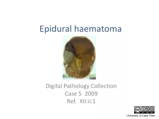
Epidural haematoma Digital Pathology Collection Case 5 2009 Ref. XII:ii:1
This is an example of a traumatic vascular injury in the central nervous system. • Unfortunately there is no clinical data for the specimen.
Click here for 3D view
A side view of the specimen shows a fracture in the left parietal region.
This is a common site for a skull fracture. • The pterion is the weakest part of the skull http://en.wikipedia.org/wiki/Pterion • The anterior branch of the middle meningeal artery runs through the dura beneath the pterion.
The skull fracture has resulted in laceration of artery, • followed by a massive arterial haemorrhage. • The haematoma which formed has a smooth convex profile. • The haematoma would have compressed the underlying brain, resulting in pressure effects leading to the patients death.
On the opposite undamaged side of our specimen the pterion can be seen as a thin, almost transparent area of skull. • The grooves made by branches of the middle meningeal are seen on the inner skull in the same region • (The grooves of the anterior and posterior meningeal arteries can also be seen.)
Inner skull • Note the location of the haematoma, it lies between the dura and the internal surface of the skull i.e. it is epidural or extradural . • (Under arterial pressure the expanding haematoma stripped the dura from the periosteum.) • Contrast this with a subdural haematoma, which would collect between the dura and the surface of the brain haematoma dura
An epidural haemorrhage can expand rapidly and is a neurosurgical emergency. • Decompression is required, usually by craniotomy.
Epidural and subdural haemorhages are both extra-axial bleeds, occurring outside of the brain substance. • Subdural haemorrhage differs from epidural haemorrhage, with regard to • The clinical scenario (who is at risk?) • the source of the haemorrhage (what vessels bleed?) • The clinical and pathological progression of the bleed.
See these cases of subdural haemorrhage in the collection……
A selection of cases from the Digital Pathology Collection by the Department of Clinical Laboratory Sciences University of Cape Town is licensed under a Creative Commons Attribution-NonCommercial-ShareAlike 2.5 South Africa Licence The full Digital Pathology Collection is accessible at www.digitalpathology.uct.ac.za If you would like to use an image or other item from our site that is not labelled with the Creative Commons Licence Logo, please contact the curator for permission.