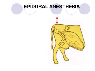
Epidural Anaesthesia
E N D
Presentation Transcript
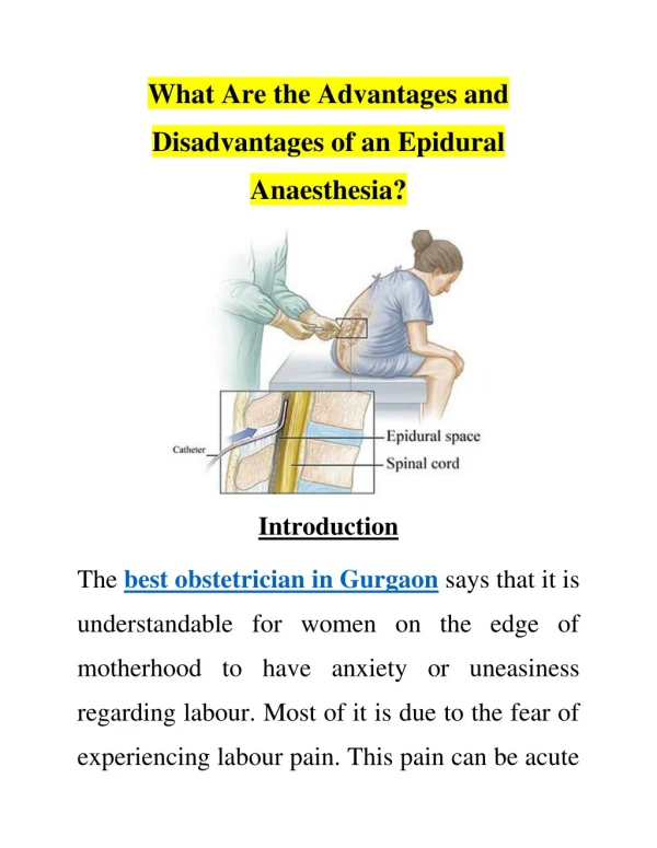
Introduction • Epidural anaesthesia is a central neuraxial block technique with many applications. The epidural space was first described by Corning in 1901, and Fidel Pages first used epidural anaesthesia in humans in 1921. In 1945 Tuohy introduced the needle which is still most commonly used for epidural anaesthesia. Its versatility means it can be used as an anaesthetic, as an analgesic adjuvant to general anaesthesia, and for postoperative analgesia in procedures involving the lower limbs, perineum, pelvis, abdomen and thorax.
Indications • Epidural anaesthesia can be used as sole anaesthetic for procedures involving the lower limbs, pelvis, perineum and lower abdomen. It is possible to perform upper abdominal and thoracic procedures under epidural anaesthesia alone, but the height of block required, with its attendant side effects, make it difficult to avoid significant patient discomfort and risk. The advantage of epidural over spinal anaesthesia is the ability to maintain continuous anaesthesia after placement of an epidural catheter, thus making it suitable for procedures of long duration
Contraindications • SAME AS SPINAL ANESTHESIA
Technique of Epidural Anaesthesia • Using local anaesthetic raise a subcutaneous wheal at the midpoint between two adjacent vertebrae. Inflitrate deeper in the midline and paraspinously to anaesthetise the posterior structures. • Insert epidural needle to the skin at this point, and advance through the supraspinous ligament, with the needle pointing in a slightly cephalad direction. Then advance the needle into the interspinous ligament, which is encountered at a depth of 2-3 cm.until distinct sensation of increased resistance is felt as the needle passes .
With 5-10ml of air in the syringe, attach it to the hub of the needle once it has entered the interspinous ligament. The plunger is gently pressed, and if there is resistance ("bounce"), the needle is very carefully advanced, with the dorsum of both hands resting against the back to provide stability. After 2-3mm, the plunger is again gently pressed, and this procedure is repeated as the needle is carefully advanced through the tissues. The distinctive decrease in resistance when the needle enters the ligamentumflavum is felt, and the process is continued in 2mm increments. There is usually a distinctive "click" when the needle enters the epidural space, and provided great care is taken, and the needle only advanced in 2mm increments, the needle should stop before it reaches the dura. At this point air can be injected into the epidural space very easily. The syringe is removed and the catheter threaded as below.
Remove the syringe and thread the catheter gently via the needle into the epidural space. The catheter has markings showing the distance from its tip, and should be advanced to 15-18cm at the hub of the needle, to ensure that a sufficient length of catheter has entered the epidural space. Remove the needle carefully, ensuring that the catheter is not drawn back with it. The markings on the needle will show the depth of the needle from the skin to the epidural space, and this distance will help determine the depth to which the catheter should be inserted at the skin. For example, if the needle entered the epidural space at a depth of 5cm, the catheter should be withdrawn so that the 10cm mark is at the skin, thus leaving approximately 5cm of the catheter inside the epidural space, which is an appropriate length.
Choice of drugs • The choice of drugs administered epidurally depends on the indication for the epidural: • Surgical anaesthesia - requires dense sensory block and usually moderate to dense motor block. To achieve this, concentrated local anaesthetic preparations are required. The most commonly used local anaesthetics in this setting are 2% lignocaine 10-20ml (with or without adrenaline 1:200 000) or 0.5% bupivacaine 10-20ml. The latter has a longer duration of action, but a slower onset time, compared with lignocaine. • For analgesia during labour, 0.1-0.25% bupivacaine 5-10ml is more popular, as it produces less motor block. • Postoperative analgesia, weaker concentrations of bupivacaine, e.g. 0.1-0.166% with or without added low dose opioids.
Complications and Side Effects • Hypotension • Inadvertent high epidural block • Local anaesthetic toxicity • Total spinal
Management of total spinal • Airway - secure airway and administer 100% oxygen • Breathing - ventilate by facemask and intubate. • Circulation - treat with i/v fluids and vasopressor. • Continue to ventilate until the block wears off (2 - 4 hours) • As the block recedes the patient will begin recovering consciousness followed by breathing and then movement of the arms and finally legs. Consider some sedation (diazepam 5 - 10mg i/v) when the patient begins to recover consciousness but is still intubated and requiring ventilation. • Accidental dural puncture is usually easily recognised by the immediate loss of CSF.