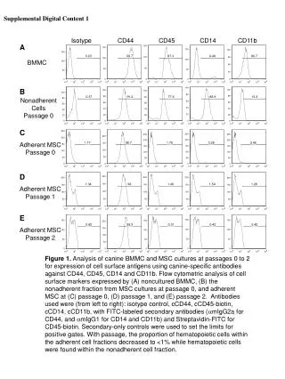
ISOTYPE SWITCH
ISOTYPE SWITCH. C C δ C3. C 1 C ε 2 C1. C 1 C 4 C ε 1 C 2. C C δ. Embryonal DNA. Somatic recombination D – J. Rearranged DNA. Somatic recombination V – D – J. C C δ. Primer RNA transcript. Transcription. Ig ISOTYPES C µ IgM C γ 1 IgG C γ 2 IgG

ISOTYPE SWITCH
E N D
Presentation Transcript
C Cδ C3 C1 Cε2 C1 C 1 C4Cε1 C2 C Cδ Embryonal DNA Somatic recombination D – J Rearranged DNA Somatic recombination V – D – J C Cδ Primer RNA transcript Transcription Ig ISOTYPES Cµ IgM Cγ1 IgG Cγ2 IgG Cγ3 IgG Cγ4 IgG Cα IgA Cε IgE C Cδ Processing C mRNA Translation Nascent polypeptide C Modification Heavy chain IgM
Cm Cd Cg3 Cg1 Ca1 Cg2 Cg4 Ce Ca2 Sm Sg3 Sg1 Sa1 Sg2 Sg4 Se Sa2 Switch regions • Upstream of C regions are repetitive regions of DNA called switch regions. (The exception is the Cd region that has no switch region). • The Sm consists of 150 repeats of [(GAGCT)n(GGGGGT)] where n is between 3 and 7. • Switching is mechanistically similar in may ways to V(D)J recombination. • Isotype switching does not take place in the bone marrow, however, and it will only occur after B cell activation by antigen and interactions with T cells.
Cm Cd Cg3 Cg1 Ca1 Cg2 Cg4 Ce Ca2 Sg3 Cd Cd Cg3 Cm Cm Sg1 Cg3 Cg1 V23D5J4 V23D5J4 Cg3 V23D5J4 Ca1 Ca1 IgG3 produced. Switch from IgM IgA1 produced. Switch from IgM IgA1 produced. Switch from IgG3 V23D5J4 V23D5J4 V23D5J4 Cg3 Ca1 Ca1 Switch recombination At each recombination constant regions are deleted from the genome An IgE - secreting B cell will never be able to switch to IgM, IgD, IgG1-4 or IgA1
Organisation of the functional human heavy chain C region genes Cm Cd Cg3 Cg1 Ca1 Cg2 Cg4 Ce Ca2 J regions Antibody isotype switching Throughout the immune response the specificity of an antibody will be essentially the same (notwithstanding affinity maturation) The effector function of antibodies throughout a response needs to change drastically as the response progresses. Antibodies are able to retain Variable regions whilst exchanging Constant regions that contain the structures that interact with cells.
CCδ C2 C4 C C Rearranged DNA in IgM-producing cell Switch regions C Cδ, C2, C4 ISOTYPE SWITCH All isotype switch recombination is productive Different recombinationsignal sequences and enzymes from VDJ rearrangement Happens after antigenic stimulation Regulated by external signals, not random C C Rearranged DNA in IgE-producing cell Primary RNA transcript C mRNA Hyper IgM syndrome Type 2. Activation Induced Cytidine Deaminase RNS editing enzyme NO HYPERMUTATION AND ISOTYPE SWITCH -Heavy chain
VL J2 gene product V35 gene product CDR1 CDR2 CDR3 Complementary Determining Region = hypervariable region
HVR3 150 Variability Index 100 HVR2 HVR1 50 FR2 FR1 FR4 FR3 0 25 75 100 50 Amino acid residue STRUCTURE OF THE VARIABLE REGION • Hypervariable (HVR) or complimentarity determining regions (CDR) • Framework regions (FR)
SOMATIC HYPERMUTATION Day 0. Ag Plasma cell clones 1 2 3 4 5 6 7 8 Day 7 PRIMARY immune response AFFINITYMATURATION 9 1011 12 13 14 15 16 Day 14 Day 14. Ag 17 1819 20 21 22 23 24 Day 21 SECONDARY Immune response Hypervariable regions
Day 6 Day 12 Day 8 Day 18 Clone 1 Clone 2 Clone 3 Clone 4 Clone 5 Clone 6 Clone 7 Clone 8 Clone 9 Clone 10 Deleterious mutation CDR1 CDR2 CDR3 CDR1 CDR2 CDR3 CDR1 CDR2 CDR3 CDR1 CDR2 CDR3 Beneficial mutation Neutral mutation Somatic hypermutation leads to affinity maturation Lower affinity - Not clonally selected Higher affinity - Clonally selected Identical affinity - No influence on clonal selection Hypermutation occurs under the influence of activated T cells Mutations are focussed on ‘hot spots’ (i.e. the CDRs) and are due to double stranded breaks repaired by an error prone DNA repair enzyme.
ANTIBODY MEDIATED EFFECTOR FUNCTIONS • Neutralization – binding of the antibody inhibits the binding of the pathogen to the cell surface, entry to the cell or multiplication • Opsonization – binding of the antibody triggers complement activation and binding to the cell surface by complement (CR1) and IgG (FcR) receptors • Cytophylic property - antibody isotypes have distinct complement activating and FcR binding activity
COMPLEMENT ACTIVATION OPSONIZATION FcR CR PHAGOCYTOSIS DEGRADATION Macrophage Ig Fc region Conformational change? Association? COMPLEMENT ACTIVATION – classical pathway BINDING TO CELLS – cytophilic property SECRETED ANTIBODIES BIND TO THE ANTIGEN IMMUNE COMPLEX ISOTYPE DEPENDENT IgG1 and IgG3 >> IgG2 és IgG4
ANTIBODY MEDIATED EFFECTOR FUNCTIONS SPECIFIC ANTIBODY Bacterium in the interstitium Bacterial toxin Bacterium in the plasma Toxin receptor Neutralization Opsonization Complement activation COMPLEMENT Internalization Phagocytosis Phagocytosis and lysis
OPSONIZATION Binding of antibody increases phagocytosis FcR COMPLEMENT ACTIVATION Opsonization by C3b PLAZMA CELL Complement C3b FcR FcR CR1 EFFECTOR FUNCTIONS OF ANTIBODIES INHIBITION Binding of bacteria to epithelial cells Binding of viruses to receptor Binding of bacterial toxins to target cells NEUTRALIZATION Small proportion of antibodies PHAGOCYTES ENGULFMENT, DEGRADATION
T – CELLS PROMOTE B – CELL DIFFERENTIATION ANTIGEN T-CELL CYTOKINES PLASMA CELL B -CELL ISOTYPE SWITCH AND AFFINITY MATURATION OCCURS IN COLLABORATION WITH T – CELLS ONLY HOW T – CELLS RECOGNIZE ANTIGENS?
ab V C B- AND T-CELL RECEPTORS SHARE BASIC STRUCTURE mIg H mIg L TCR T cell receptor TCR TCR TCR = + The variable region of the -chain is generated by gene rearrangements of the V – D – J gene segments analogous to the generation of IgH diversity The variable region of the -chain is generated by the recombination of V and J analogous to IgL T-CELL C Single binding site No somatic mutation
The VARIABLE REGIONS OF - AND -CHAINS ARE GENERATED BY SOMATIC RECOMBINATION Recombination of V and J genes can occur after multiple unsuccessful recombination T-CELL Antigen receptor TCR not functional a/b next funcional V C further funkcional (no allelic exclusion) mRNS
ESTIMATED VARIABILITY OF IMMUNOGLOBULIN AND T-CELL RECEPTOR GENES NO SOMATIC HYPERMUTATON
CHARACTERISTICS OF T-CELL ANTIGEN RECOGNITION ACCESSORY CELL B-CELL T-CELL ANTIGEN BINDING NO INTERACTION T-CELL ACTIVATION Antigen receptor • The TCR is not able to interact directlywith soluble or cell-bound antigen • T-cell activation can be induced by antigen in the presence of acessory cells, only • 3. T-cells recognize virus-infected cells V C a/b
TCR BCR Kinases Syk ZAP70 Btk Itk Adaptors + substrates PLCg2 PLCg1 Lyn SLP-65/BLNK SLP-76 fyn Similar but not identical signaling elements in B and T cells
T CELL RECEPTOR MEDIATED SIGNALING εδεγ αβ ζζ Multisubunit Immune Recognition Receptors MIRR ITAM Immunoreceptor Tyrosine-based Activation Motif ACTIVATION
APC T CELL
THE INTERACTION OF T CELLS AND ANTIGEN PRESENTING CELLS 1 2 3 4 5 6 7 8 interaction recognition stabilization separation Negulescu P.A. et. al. Immunity 4: 421-430, 1996
THE IMMUNOLOGICAL SYNAPSE ANTIGEN PRESENTING CELL ICAM-1 B7 CD4 CD28 LFA-1 adaptor SIGNALING COMPLEX T CELL ACTIVATED T CELL CD48 CD2 ICAM – Intercellular Adhesion Molecule
THE CONTACT OF APC AND T CELLS IS STABILIZED BY ADHESION MOLECULES * * T CELL B CELL * * * *
RECOGNITION EFFECTOR CELL Plasma cell B-lymphocyte cytokines BCR + antigen Antibody production Cytotoxic T-limfocyta (Tc) Cell killing TCR + peptide + MHC-I Effector cell retains specific receptor Effector cells secrete cytokines cytokines Helper T-lymphocyte (Th) Macrophage activation Lymphocyte activation Inflammation TCR + peptide + MHC-II
Tc Th Exogenous Ag Endogenous Ag Peptides of exogenous proteins (toxin, bacteria, allergen) bind to class II MHC molecules Peptides of endogenous proteins (virus, tumor) bind to class I MHC molecules RECOGNITION OF EXOGENOUS AND ENDOGENOUS ANTIGENES BY T-LYMPHOCYTES
Signal 1 antigen & antigenreceptor Th APC ACTIVATION Signal 2 B7 family members (CD80 & CD86) CD28 Antigen presentation - T cells are co-stimulated Costimulatory molecules are expressed by professional APC including dendritic cells, monocytes, macrophages, and B cells, but not by cells that have no immunoregulatory functions such as muscle, nerves, hepatocytes, epithelial cells etc.
ROLE OF CO-STIMULATION IN THE ACTIVATION OF HELPER T CELLS CD40L CD28 CD40 B7 B7 APC APC APC NORMAL TISSUE CELLS DO NOT EXPRESS CD40 OR B7 CO-STIMULATORY MOLECULES
Antigen 1 IL-2 IL-2 IL-2R IL-2R Mechanism of co-stimulation in T cells Resting T cells Signal 1 NFAT binds to the promoter of of the a chain gene of the IL-2 receptor. The a chain converts the IL-2R to a high affinity form Express a low affinity IL-2 receptor- and chains and produce no IL-2
Costimulation Antigen 2 1 IL-2 IL-2R Mechanism of co-stimulation in T cells Signal 2 Activates AP-1 and NFk-B to increase IL-2 gene transcription by 3 fold Stabilises and thus increases the half-life of IL-2 mRNA by 20-30 fold IL-2 production increased by 100 fold overall Immunosuppressive drugs illustrate the importance of IL-2 in immune responses Cyclosporin & FK506 inhibit IL-2 by disrupting TcR signalling Rapamycin inhibits IL-2R signalling
IL-2 α β γ CYTOSKELETON THE HIGH AFFINITY IL-2 RECEPTOR Ligand binding No signaling JAK Janus kinase STAT Signal Transducer and Activatior of Transcription Gene transcription Proliferation
INITIATION OF T CELL PROLIFERATION IL-2R IL-2 IL-2Rα IL-2R low affinity adhesion recognition IL-2R high affinity transferrin costimulation insulin IL-1 IL-2 IL-2 AUTOCRINE GROTH FACTOR PROLIFERATION
CSA FK506 MECHANISM OF THE ACTION OF THE IMMUNE SUPPRESSIVE DRUGY CYCLOSPORIN A AND FK506/PROGRAPH TCR-CD3 Other receptors Inactive phosphatase Aktive phosphatase Dephosphorylation of cytoplasmic NF-AT induces translocation to the nucleus FKBP2 isomerase Cycliphilin A isomerase Cytokines, activation molecules IL-2, IL-2R TGF NOT ANTIGEN SPECIFIC
CO-STIMULATION IS ESSENTIAL FOR PRIMING OF NAIVE T LYMPHOCYTES The antigen-specific and the co-stimulatory signals have to be induced in concert to induce T lymphocyte activation The antigen-specific and co-stimulatory signals can be delivered simultaneously by professional antigen presenting cells, only The antigen-specific and the co-stimulatory singnals has to be delivered by the same professional antigen presenting cell
T CELLS REQUIRE TWO SIGNALS TO GET ACTIVATED APC not presenting antigen Activated APC Resting APC B7 B7 CD4 CD4 CD4 CD28 CD28 CD28 21 1 2 T-cell activation T-cell anergy No effect ANTIGEN SPECIFIC ACTIVATION, ANERGY OR NEGLECTION
Naïve T cell Antigen 1 IL-2 IL-2R Epithelial cell Anergy Signal 1only The T cell is unable to produce IL-2 and therefore is unable to proliferate or be clonally selected. Unlike immunosupressive drugs that inhibit ALL specificities of T cell, Signal 1 in the absence of signal 2 causes T cell unresponsiveness to a specific antigen Self peptide epitopes presented by a non-classical APC e.g. an epithelial cell
PROFESSIONAL ANTIGEN PRESENTING CELLS Express MHC class I and class II molecules Express co-stimulatory molecules (B7, CD40) Take up extracellular antigens B cells – soluble proteins, toxins (ADAPTIVE) Macrophages – extracellular pathogens (bacteria, yeast) INNATE – particles Dendritic cells – viruses, apoptotic cells
CHARACTERISTICS OF PROFESSIONAL ANTIGEN PRESENTING CELLS Macrophage Dendritic cell B - lymphocyte Ag uptake phagocytosis +++ phagocytosis +++ Ag-specific mIg virus infection ++++ ++++ MHC expression induced +/+++ constitutive ++++constitutive +++ bacteria, cytokine immature/mature +++/++++ activation ++++ Pesented Ag particulate Ag protein soluble protein intra/extracellular virus protein, allergen toxin pathogens apoptotic cell Co-stimulation induced +/++ constitutive ++++induced +/+++ éretlen/érett+++/++++ Localization lymphoid tissue lymphoid tissue lymphoid tissue connective tissue connective tissue peripheral blood body cavities epithelium Lymph node evenly immature –tissue follicles mature –T cell area
CHANGES IN TISSUE ENVIRONMENT RESULTS IN MACROPHAGE AND DENDRITIC CELL ACTIVATION Macrophage Monocyte Dendritic cell Activated dendritic cell Virus, extracellular pathogenes, inflammatory cytokines (LPS, TLR) DANGER SIGNAL BLOOD TISSUE LYMPH Phagocytosis of bacteria, degradation (LPS, TLR) DANGER SIGNAL Activated macrophage
ACTIVATION AND MIGRATION OF DENDRITIC CELLS Effector and memory T cells LYMPH Activated DC Inflammation Pathogen Naive T cells ANTIGEN BLOOD Tissue DC TISSUE LYMPH NODE TISSUE INTERACTION OG DC AND T CELL T CELL ACTIVATION
DENDRITIC CELL T - LYMPHOCITE INTERACTION IN THE LYMPHOID ORGANS Activated dendritic cells in the lymphatic tissues act as antigen presenting cells Tight contact with specific T cells
No danger Cell containing only self antigens APC No danger Apoptotic cell death. A natural, often useful cell death. APC The danger hypothesis & co-stimulation Full expression of T cell function and self tolerance depends upon when and where co-stimulatory molecules are expressed. Innocuous challenge to the immune system fails to activate APC and fails to activate the immune system Fuchs & Matzinger 1995
Necrotic cell death e.g. tissue damage, virus infection etc APC APC DANGER Pathogens recognised by microbial patterns The danger hypothesis APC that detect ‘danger’ signals express costimulatory molecules, activate T cells and the immune response