Download

1 / 52
520 likes | 1.08k Vues
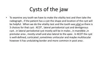
Cysts of the jaws and neck. Development of a Cyst. The common behavioral feature of all cysts is the stimulation of residual developmental epithelial cells, leading to proliferation but not invasion of adjacent tissues.

E N D
Development of a Cyst • The common behavioral feature of all cysts is the stimulation of residual developmental epithelial cells, leading to proliferation but not invasion of adjacent tissues. • The epithelial rests proliferate into a solid mass of epithelial cells. As the mass enlarges, the epithelial cells in the center become positioned further from the blood supply at the periphery of the mass. • At some point, usually the cells at the center become too far removed from the nearest blood vessel to survive by nutritional diffusion. They die, creating a lumen. • Their intracellular products make the lumen hypertonic, which transudates fluid into the lumen. • This in turn creates a hydrostatic pressure, producing bone resorption, clinical expansion, and sometimes mild paresthesia or pain.
As additional epithelial cells die off and are sloughed into the lumen, their contents perpetuate the hypertonic state and the hydrostatic pressure. • The cell membranes and nuclear membranes of these sloughed cells are high in cholesterol, hence the common finding of cholesterol clefts in the lumen or even in the walls of many cysts. • As the cyst enlarges, it compresses surrounding connective tissue into a connective tissue wall. • The epithelial lining matures and develops a basement membrane. • The cyst lining continues to proliferate, thus causing the cyst to enlarge until (1) it is removed (enucleation), (2) the proliferating cells are communicated into the oral cavity or an external surface so as to break the proliferation‐hydrostatic pressure cycle (marsupialization), or (3) the inciting cytokines are removed (via tooth removal or root canal therapy in radicular cysts).
Gingival Cyst of Newborn (Bohn’s Nodules) Etiology • Cystic degeneration of residual dental lamina/odontogenic epithelium • Found in over 80% of newborns
Clinical Presentation • Small (1–2 mm), usually multiple, yellow-white nodules over the alveolar crest in neonates • Usually involute following spontaneous cyst rupture
Diagnosis • Appearance and location. • Histologically, parakeratinized epithelial lining with keratinfilled cyst cavity is noted. Differential Diagnosis • Eruption cyst
Treatment • None; observation only Prognosis • Excellent
Odontogenic Keratocyst Etiology • A benign, aggressive developmental odontogenic cyst; may be associated with mutation of PTCH tumor suppressor gene • There is general agreement that OKCs develop from dental lamina remnants in the mandible and maxilla.
Clinical Presentation • 5 to 15% of odontogenic cysts • Usually occurs sporadically as an isolated finding • Approximately 5% are associated with nevoid basal cell carcinoma. • 5% of patients have multiple odontogenic keratocysts (OKCs) and no syndrome
Radiographic Findings • Can occur in any area of maxilla or mandible • Rarely may arise in gingival soft tissue only (peripheral) • Mandible is preferred site in 65 to 78% of cases • Often (40%) seen in a dentigerous relationship • Discrete radiolucency, usually in relation to teeth (apical, lateral radicular, pericoronal to impacted tooth) • May be unilocular to multilocular
Microscopic Findings • Thin, parakeratinized epithelial lining (6–10 cells thick) • Wavy, corrugated surface configuration • Prominent, palisaded, cuboidal to low-columnar basal cell layer • Basal layer “budding” into fibrous stroma is seen occasionally • Satellite or daughter cyst formation noted frequently
Diagnosis • Radiographic features • Microscopic findings Differential Diagnosis • Odontogenic cysts: dentigerous, radicular, lateral periodontal, or glandular odontogenic • Nonodontogenic cyst: nasopalatine duct • Odontogenic tumors: ameloblastoma, myxoma • Giant cell granuloma • Central mucoepidermoid carcinoma
Treatment • Excision with curettage of bony confines. Prognosis • The recurrence rate varies from 10 to 30% (solitary OKCs). • Recurrence rates are greatest in patients with a syndrome.
Nevoid Basal Cell Carcinoma Syndrome Etiology • Autosomal-dominant condition • Loss of heterozygosity at chromosome 9q22.3 • Mutation of PTCH tumor suppressor gene
Clinical Presentation • Multiple jaw cysts (odontogenic keratocysts) • Numerous cutaneous basal cell carcinomas, which arise early in life and are independent of sun exposure • Bifid ribs • Calcification of falx cerebri • Ocular hypertelorism • Mandibular prognathism • Broad nasal bridge • Medulloblastoma • Palmar and plantar pits
Radiographic Findings • Multiple jaw radiolucencies • Lamellar calcification of falx cerebri • Bifid rib on abdominal radiograph
Diagnosis • Radiographic and clinical findings Differential Diagnosis • Other syndromes, such as the following: • Charcot-Marie syndrome • Waardenburg’s syndrome
Treatment • Excision of basal cell carcinomas and odontogenic keratocysts • Excision of other related aggressive tumors at other sites • Genetic counseling Prognosis • Guarded
Dentigerous Cyst Etiology • A developmental odontogenic cyst arising subsequent to separation between dental follicle and the crown of an associated unerupted tooth • Proliferation of reduced enamel epithelium lining the follicle, with fluid accumulation between epithelium and impacted tooth crown • Alternatively, degeneration of the stellate reticulum component of the enamel organ occurs during odontogenesis.
Clinical Presentation • Most commonly involves frequently impacted teeth: mandibular third molars, followed by maxillary canines • Usually noted during second and third decades • Asymptomatic; discovered on routine radiographic examination • Painless jaw/alveolar expansion may occur; cortex is thinned and rarely perforated
Radiographic Findings • Well-defined radiolucency enclosing crown of unerupted tooth • Corticated/opaque margins unless infected • May produce root resorption of adjacent erupted teeth • Usually unilocular; less commonly multilocular
Diagnosis: Microscopic • Cysts without secondary inflammation • Thin, cuboidal, nonkeratinized epithelial lining two cell layers thick with flat epithelial–connective tissue interface • Loosely arranged collagen bundles, occasionally containing scattered odontogenic epithelial rests • Cysts with secondary inflammation • Hyperplastic, nonkeratinized squamous epithelial lining with epithelial ridge development • Variable chronic inflammatory cell infiltrate within condensed collagen stroma
Differential Diagnosis: Radiographic • Odontogenic keratocyst • Ameloblastoma Treatment • Cyst enucleation and extraction of associated tooth • Marsupialization prior to excision may be considered if the cyst is very large. Prognosis • Excellent • Possible complications • Pathologic fracture with large lesions • Neoplastic transformation of epithelial lining (Ameloblastoma and, rarely, squamous cell carcinoma)
Eruption Cyst Etiology • Soft tissue cyst of attached gingiva secondary to fluid accumulation within the follicular space of an unerupted tooth Clinical Presentation • Gingival swelling on the alveolar crest • Usually soft, translucent to bluish (“eruption hematoma”)
Diagnosis • Location • Radiographic demonstration of erupting tooth Differential Diagnosis • Gingival cyst
Treatment • Usually none is necessary as tooth typically erupts through lesion • Possibly unroof cyst to facilitate eruption Prognosis • Excellent
Lateral Periodontal Cyst Etiology • Stimulus unknown • Dental lamina remnant proliferation within the alveolar segment of the jaw, separate from the periodontal ligament
Clinical Presentation • Asymptomatic • Usually occurs in fourth decade and beyond • Usually in mandibular canine/ premolar region (65%) • In the maxilla, the lateral incisor area predominates.
Radiographic Findings • Well delineated, round to ovoid lucency with thin, opaque (corticated) margin • Located lateral to vital tooth roots • Usually unilocular; may be multilocular (botryoid odontogenic cyst)
Diagnosis • Thin, nonkeratinized epithelial lining • Nodular epithelial thickening along cyst lining • Lining cells are cuboidal with interspersed clear glycogen-filled cells. Differential Diagnosis • Inflammatory, lateral radicular cyst • Primordial cyst/odontogenic keratocyst • Odontogenic tumor • Glandular odontogenic cyst
Treatment • Conservative enucleation • The botryoid variant requires more aggressive curettage. Prognosis • • Recurrence uncommon • • Increased risk of recurrence with botryoid variant; longer-term follow-up necessary
Radicular Cyst Etiology • Preceded by periapical granuloma; arises as follows: • Secondary to necrosis of dental pulpal tissue. • Stimulation of epithelial network (Malassez’s rest) at tooth root apex results in cystification. • Cyst growth continues secondary to effects of osmotic gradient across epithelial lining layers, mediators of inflammation, and epithelial proliferation.
Clinical Presentation • Asymptomatic unless there is an acute exacerbation • Usually a limited process at root apex or lateral to root surface • Radiograph shows a round and well-defined lucency, usually with a sclerotic margin. • Generally 1 cm or less across, but can be significant in size • Root resorption uncommon
Microscopic Findings • Stratified squamous epithelial lining • Lumen filled with cell debris, fluid, cholesterol • Connective tissue wall with mixed inflammatory infiltrate
Diagnosis • Documentation of nonvital tooth • Radiograph shows alteration of apical bone Differential Diagnosis • Periapical granuloma • Central giant cell granuloma • Odontogenic and nonodontogenic tumors • Metastatic tumor
Treatment • Endodontic therapy or • Periapical surgery and biopsy or • Tooth extraction and biopsy Prognosis • Excellent • Occasional recurrences