Download
1 / 26

E N D
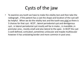
Cysts of the jaw • To examine any tooth we have to make the vitality test and then take the radiograph , if the patient has a cyst the shape and location of the cyst will be helpful . When we do the vitality test and the tooth was vital so there is 3 choices for that cyst : KCOT , lateral periodontal cyst and dentigerous cyst , in lateral periodontal cyst mostly will be in males , in mandible ,in premolar area , mostly small and also lateral to the apex . In KCOT the cyst is well-defined, corticated ,sometimes unilocular and maybe multilocular however it has undulating border and more common in post area .
3)lateral radicular cyst 2)Residual cyst 1)Apical radicular cyst * Radicularcyst : is 3 types apical , residual and lateral radicular ,all of these the origin is tooth so they are Odontogenic cysts and the tooth is non vital . radiographically they are radiolucent. I-If the cyst attached to the tooth then the tooth is non vital otherwise the tooth will be vital
-Early radicular cyst ,, epithelium is irregular , variable in thickness .wall is vascular and chronic inflammatory cell infiltrate (CICI) can be seen .
*Late radicular cyst : it will happen if the pateint didn’t go to the dr. to cure the cyst or the dr. didn’t repair it perfectly . • epithelium will be more unifom and the wall will be more fibrous and less vascularity , Rushton bodies (hyalinizedmaterial from epithelial product ) can be seen as well .
Dentigerous cyst : its attached to the cementoenamel junction ,covering the crown of the uneruptedteeth , well definied ,corticated ,unilocular and has typical cemtoenamel to cementoenamel junction ,, there are lateral ,central and circumferential types
histopathology of the dentigerous cyst : cuboidalepithelium ,thin 2-4 layers, has a mucus cells and fibrous cells but theres NO INFLAMMATORY EXUDATE (because this cyst is developmental not inflammatory ) although sometimes it may have inflammatory infltrate because there is communication with the outside so there will be fibrobalst and collagen bundle every where .
lateral periodontal cyst : tear drop in shape ,typical lateral between the root , originated from rest cells of serres . varians of this cyst is Botryoidodontogenic cyst and its multilocular so its more aggressive
Buccal bifurcation cyst : on the buccal surface of the bifurcation,, to distinguish between buccal bifurcation cyst (that is variant of the paradental that is variant of the dentigerous cyst ) and radicular variant cyst by vitality test ,, if the tooth is vital and the cyst bilaterally so its bifurcation cyst while if the tooth is non-vital then its radicular variant cyst.
gingival cyst : its looks like lateral periodontal cyst happened in the soft tissue , most commonly seen in newborn infants , if its on the midline they called it epstein pearls because it appears white in color . Histopatology :appears like lateral periodontal cyst ,its thin ,unifom with no or minimal inflammatory cells and the special feaure is the plaques (the thickest area in the epithelium ).
keratocysticodontogenic tumor (KCOT) : the cells has inherent ability to replicate and its name tumor is fair because the cells are replicating and its treatment is close to tumor not to the cyst . local recurrence is high and we remove it surgically ,,the problems in this treatment way is : 1)epithelial lining is not attached to the submucosarather there is a flat junction so the epithelium might detach and the surgeon might leave some and these are capable of forming a new cyst .
- This picture shows a hitological section of a KCOT , we can see a lining epithelial that is not that thick (6-10 layers ) , we have Palisadedbasal cells , and we have a flat junction between the epithelium and the submucosa .The basal cells here they are psudocolumnar not columnar but because they are palisading , they give you that look (as if they were columnar but they are not columnar)
This picture shows Epithelial residues and Satellite ( daughter cysts ) • -The cyst normally have a space then the epithelial lining then submucosa , but in KCOT we then see epithelium again and this is an abnormal thing to see in a typical cyst , so if the surgeon removed the cyst without removing the satellite cysts and reaching what so called “safety margin “, then their epithelium is capable of forming new cyst, so sometime surgeons use different chemical substances that kills the residual cysts to make sure that there will be no recurrence and that no more cysts will appear in that place.
- Epithilial budding , is another thing that can be seen in KCOT , and again the surgeon has to make sure that he removes all that epithelium as it can form a new cyst if it remains there .
- All what we have been talking about are Odontogenic cysts , now we will talk about non odontogenic cysts “it has nothing to do with the presenting teeth nor with the teeth during development so they don’t arise form – epithelial cell rest of Malassez or what ever or epithelial residual or from infected tooth caused the epithelial to replicate.
-Incisive Canal Cyst “Nasopalatine duct cyst “ ,on radiograph , it has a heart shape.- The Incisive canal “duct “is normal anatomy that has an opening of a certain size, and if for some reason ( a developmental one not environmental ) it will become bigger in size , and here we say that on a radiograph we see the incisive foramen and notice two things is it growing bigger ( bigger than 8 mm )? , is it affecting the adjacent teeth ?
- some times the foramen is normally lager but not larger than 8 mm ( pathologist say 6 mm and radiologist say 8 mm so to be on the safe side we say 8 mm ) , if its larger than that then that’s probably means it is a cystic degeneration . - The other thing is that if we notice that the adjacent teeth are affected ( by root resorption or being displaced ) this indicate that it is a cyst , because a normal anatomic structure wont do such a thing . -This cyst is treated by excision , it has no recurrence rate .
- a Picture showing an MRI image using non ionized radiation forNasopalatine cyst , and its frequency makes any fluid looks white in color – yelma3- .. Here we can see that the incisive duct cyst is fluid filled cavity like any cyst and present inside the incisive canal .- This cyst originates from the epithelial that lines the incisive canal it self not from a tooth– not odontogenic – and because this canal connect between the nose and the oral cavity so the oral part of the cell has more sequamous cell and keratinized while from the nasal part it has more respiratory epitheluim “ciliated pseudostratifiedcolumnar epithelium” so its more columnar and has mucus cells as will. • -
-Solitary bone cyst or Traumatic bone cyst , this is a Pseudocystcause under the microscope and when we examine the histological sections we find that it does not have an epithelial lining , and its caused by trauma .- when we say trauma it does not necessarily means that trauma is from a car accident , it can be less severe like eating hard .stuffs and such – These cysts are very typical , seen in the young age patients (10-20 yrs).
-what actually causes the cyst is that when we have a trauma we will have bleeding and then organization by fibrosis and then healing by new bone formation , but if that blood clot integrates for a reason or another then the space will stay empty and it will take much longer time to heal than when the normal healing process takes its place . • - If we open theses cysts or try to take an aspiration by a needle from them ,then usually you will not find anything , its just an empty cavity .
- we have two schools for the treatment of these cysts , one says that you can’t be 100% sure that’s it’s a traumatic cyst so you have to open it , you see that’s is empty then u know it’s a traumatic , you scratch its wall and fill it with blood and close it , by that you allow the normal healing process to take place , so – the etiology is trauma and the treatment is also trauma.- the other school says that if from the x-ray you are so sure that’s is a traumatic bone cyst , and you know that your patient will come later to you – ma ra7 yroo7 3l mozambee8 xD – and you will be able to follow up that patient , then you don’t do anything except waiting and follow up , cause they say that what happened –trauma- is going to delay the healing process rather than stopping or preventing it , so instead of healing in one year its going to take maybe 3 yrs . - However , most drs , would actually chose to go in and see what's in that cyst because it is really hard to make sure that’s it a traumatic bone cyst depending on a radiograph only , and because our patients most probably will not come back for follow up and also because the same radiographic appearance might be KCOT in its early stages where there is no bone resorption or displacement yet . - The size of this cyst does not increase rather it decreases if we waited long enough , and its Painless , and its just an empty cavity that does not affect the bone – resorption – nor the teeth – resorption and displacment-
- This x-ray shows Stafnes’s idiopathic bone defect, is not actually a cyst although its in the mandible and uniloculer and corticated but its not a cyst .- you’re going to have a hard time to know whether it is buccal or lingual or centered.- From this radiograph we can take two infromations , 1- its non- odontogenic cause its away from the tooth beneath the inferioeralveoler canal , 2- its cortical border is thicker than usual – el mafroodtkoonzy 2alam mabry w torsomfe el border bs hay a3rad mnhaik -
- when they take a 3D image they find that it’s actually a concavity on the mandible lingual surface and this concavity has a salivary gland tissue in it . - The etiology of this ectopic salivary gland is unknown , we have lots of theory , but no body really knows .-It can be an extra salivary tissue from the sub mandibuler gland or the parotid gland or sub lingual gland , and this tissue sets on a concavity on the lingual surface of the mandible .
- This anatomic concavity “Stafne’s bone defect “ appears radiolucent on a x-ray as a cyst looks , but the reason is actually because having a cavity will make the mandible thinner there and so it will not attenuate as much as the thicker normal non-cavitated mandible so it will let more photons pass so it appears black – radiolucent –- The reason that makes the cortex thicker than a normal cyst , because this is a the cortex of the mandible it self not cortex of a lesion.
- sometime the stafne’s bone defect is found anteriorly , and it can be confusing , you might think of doing a vitality test then u will find that all teeth are vital , and then you think it does not look like a KCOT because its very well defined and round , and its not lateral periodontal cyst – la2enha wa6yeh kteer - , so you will ask for a 3D image and discover that’s it’s a cave – concavity – anteriorly , now those are rare and not as common as the submandibuler , those are for the sub lingual gland .- We have stafne’s cyst for the parotid gland in the ramus of the mandible , it can be confuse with many lesions that happen in the ramus . - In general , when a lesion is found up in the ramus, you always would like to do a 3D imaging before you open the lesion and discover that – Oops ! Fotnabel parotid –- Stafne’s bone defect are developmental and are considered as pseudocyst , you don’t need to do anything for the patient since it’s a salivary gland , only document that in the patient’s record so that the next dentist wont be confused again. ** el mafroodykoonfepiclalsublingal gland Stafne’s cyst bs ma la2ait bel net weldr ma a36alt slidat-ha .