Download
1 / 21
220 likes | 339 Vues
This research explores transverse sacral fractures, their diagnostic dilemmas, and therapeutic considerations, shedding light on this unusual injury type. Learn about epidemiology, classification, and radiological difficulties in detecting TSFs.

E N D
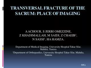
TRANSVERSAL FRACTURE OF THE SACRUM: PLACE OF IMAGING A ACHOUR, S JERBI OMEZZINE, Z KHADIMALLAH, M SAIDI, Z CHAEIB1, N SASSI1, HA HAMZA. Department of Medical Imaging, University Hospital TaharSfar, Mahdia, Tunisia 1Department of Orthopaedics, University Hospital TaharSfar, Mahdia, Tunisia MK2
Introduction -Transverse sacral fractures (TSFs) are an uncommon type of sacral fractures. -They are classified as zone III sacral fractures. -Sacral fractures are injuries that frequently are overlooked. -They present diagnostic and therapeutic dilemmas to the clinicians who evaluate trauma patients. -Between 40% and 50% of sacral fractures have a concomitant pelvicfracture.
-Usually associated with neurologic impairment and injuries of the pelvic ring -Most commonly, sacral fractures are longitudinal. -Transverse fractures of the sacrum are frequently difficult to appreciate on conventionalradiographs. -Knowledge of the range of radiological appearances of sacral fractures and when to use the appropriate imaging techniques will enable the radiologist to properly assess the sacrum.
Objectif Reports on sacral fractures, particularly transverse fractures, appear infrequently in the literature in comparison to those on other fractures of the pelvis. This may be due in part to failure to recognize them due to low suspicion of a fracture or difficult radiological examination. Reported is the case of a patient who sustained a transverse sacral fracture withsubsequentneurologiccomplications.
Materials and methods -A 20 year-old woman slipped and fell directly onto her low back. -She presented later that day with complaints of nonradiating low back pain made worse with ambulation and weight bearing. She had urinary hesitancy and could void only small amounts of urine at a time. -She denied hematuria, dysuria, or any urinary or fecal incontinence. --She sustained no other trauma in the fall.
- On examination the patient was in moderate pain. - The sacrococcygeal area was tender to palpation. Rectal examination was normal with good sphincter tone. -She demonstrated good muscle strength of the lower extremities and her sensation was intapt. -Both patellar and ankle reflexes were intact bilaterally. -The patient's pain worsened with walking.
Results • -The initial radiographs of the lumbosacral and coccygeal • areas were read as a fracture through the junction of the mid and distal thirds of the sacrum with slight anterior displacement of the distal fragment. • -Tomograms confirmed a fracture through the S 4 level with minimal displacement and angulation.
Discussion Epidemiology: -TSF are rare. -They constitute less than 1% of all spinal fractures and only 3% to 5% of all sacral fractures . -TSF are most common among young people between the second and third life decade, and are slightly more frequent in males. -There are diverse types of accidents causing TSF, almost 37% are caused by motor vehicle accidents (MVA); 35% are caused by a fall usually landing on buttocks.
Classification: * Zone I fractures involvethe alar region. *Zone II fractures occur in the sacral foraminal area. *Zone III fractures occur in the vicinity of the central canal. -Because of the location of zone III fractures, they have a high incidence of neurological deficits. -Transverse sacral fractures (TSFs) have been traditionally included as a type of zone III fractures
Diagnosis Because of the low incidence and the radiological difficulties to visualize the fracture, the diagnosis of TSF is oftendelayed. Clinicaldiagnosis: -The saddle anesthesia, loss of bladder function, and rectal tone presented in patients with sacral fractures may be masked orrunrecognized during the acute phase of trauma. -Sacral injury should be suspected in any patient reporting sacrococcigealpain . -Lacerations, bruising, tenderness, swelling, and crepitus over sacral area are signs of potential injury .
-Patients with a suspected sacral fracture must have assessment of the lower sacral roots. -Rectal examination is very important in these patients; diminished anal sphincter tone may be present even without bowel or bladder symptoms. -The neurological deficit secondary to TSF most commonly described in the literature are BBD and saddle anesthesia. -The mostcommonneurologicaldeficit was BBD characterized by incontinence, retention, or flaccid sphincters. Another common presentation was nerve root disturbance, usually L5 or S1 roots.
Radiologicaldiagnosis -Conventional radiography of the sacrum is often difficult to interpret as overlying bowel gas, bladder, and the normal angulation of the sacrum make the diagnosis of injury problematic. -The fracture does not project well on the anteroposteriorview because of the sacral orientation. -Pelvic inlet and outlet views are recommended as additional studies to improve visualization of the sacrum in any patient with a suspected sacral fracture.
-Certain findings intimate the presence of sacral fractures and suggests the need for more extensive investigation: 1)fracture of a lower lumbar transverse process; 2) pelvic ring fractures known to be associated with a sacral fracture (eg, bilateral rami fractures); 3) discontinuity or asymmetry in the ‘‘sacral notch’’; 4) irregularity of the arcuate lines in the upper three sacral foramina. -Although TSF are seen best in lateralradiographs, anteroposteriorradiographscanshow the step ladder sign caused by displacement and overriding of the fracture fragments, this finding is very suggestive of TSF.
CT SCAN • -Computed tomography is the preferred modality for diagnosing suspected or known posterior injury of the pelvic ring. • -Computed tomography with 1- to 2-mm cuts as well as sagittal and coronal reformatted views offers superior visualization of the fractured sacrum.
MRI • -Because the termination of the thecal sac at the S1–S2 interspace, myelography is of limited usefulness. Sacral magnetic resonance imaging may be useful for patients presenting with posttraumatic sacral neurological deficits which are not explained by conventional radiological test findings; however, it has little usefulness to define a fracture in an acutely traumatized patient. • -With MR imaging, the sacral plexus and surrounding structunes • can be studied in detail . • -Mubtiplanar examination aids in the assessment of the effect of diseases on the sacral plexus. The anatomic axial and oblique coronal planes display the relevant structures somewhat more distinctly than the sagittal plane does.
-Obtaining axial and oblique coronal images, with T1weighting in one plane and T2 weighting in the other, appears to be a reasonable approach to MR imaging of the sacral plexus. -If necessary, supplemental sagittal images on additional pulse sequences can be added.
Treatment -Different types of treatmentsfor TSF have been reported. -These treatments include : -conservative management -initial conservative management followed by surgical treatment -surgical treatment
Conclusion -TSF are a special type of sacral fractures which can be difficultto diagnose initially. -The U-shaped sacral fracture can be overlooked without appropriate imaging. -Although it is difficult to discern on anteroposterior radiographs and axial or coronal CT, the fracture is easily identifiable on CT images in the sagittal plane. -We advocate reconstruction of CT images of the sacrum in the sagittal plane in trauma to prevent failureof identification.