Immunohistochemistry
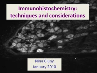
Immunohistochemistry. Introduction. Immunohistochemistry (IHC) combines histological, immunological and biochemical techniques for the identification of specific tissue components by means of a specific antigen/antibody reaction tagged with a visible label.


Immunohistochemistry
E N D
Presentation Transcript
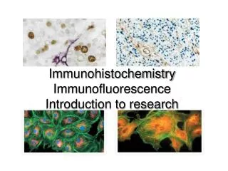
Introduction • Immunohistochemistry (IHC) combines histological, immunological and biochemical techniques for the identification of specific tissue components by means of a specific antigen/antibody reaction tagged with a visible label. • IHC makes it possible to visualize the distribution and localization of specific cellular components within a cell or tissue.
IHC is an application of antibodies to tissue preparation for the localization of target antigens: • Wide range of specific antibodies • Highly sensitive detection system
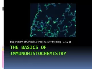
Immunohistochemistry utilizes labeled antibodies to localize specific cell and tissue antigens, and is among the most sensitive and specific histochemical techniques. • Because many targeted antigens are proteins whose structure might be altered by fixation and clearing, so frozen sections are commonly used. • In some cases, paraffin wax can be used for embedding.
Cells grown as a monolayer Cells grown, spun into a pellet, frozen or paraffin embedded and sectioned Sections from tissues contain many different kinds of cells as well as extra-cellular matrix components Immunohistochemistry assays may use cells on slides OR use tissue sections that are frozen or paraffin embedded
Tissue section on glass slide: Frozen If the tissue is frozen The sections may need to be used in immunohisto-assays as Unfixed: Advantage: antigens are unaltered Disadvantage: sections may fall off slide during staining Acetone fixed: - precipitates proteins onto cell surface---may extract lipids - is needed for many of the “CD” antibodies Paraformaldehyde fixed: - needs to be freshly made, or frozen soon after
Tissue section: Paraffin embedded If the tissue is paraffin embedded - Deparaffinize ( remove the infiltrated paraffin wax, by using organic solvents) - The section then needs to be rehydrated, by sequential immersion in graded alcohols (100%, 70% , 50% and then PBS) - The deparaffinized section may need to be treated to expose buried antigenic epitopes with either proteases or by heating in low pH citrate buffer , or high pH EDTA buffer (Antigen Retrieval)
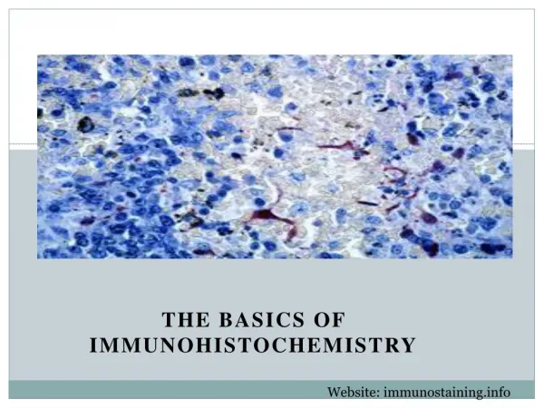
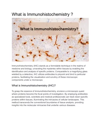
Principle • The principle of immunohistochemistry is to localize antigens in tissue sections by the use of labeled antibodies as specific reagents through antigen-antibody interactions that are visualized by a marker such as fluorescent dye, enzyme, radioactive element or colloidal gold.
Antibodies (Immunoglobulins) • Glycoprotein that are produced by plasma cells and used by the immune system to identify and neutralise foreign objects, ie. bacteria and viruses • Recognise a specific Antigen- mainly proteins, glycoprotein, polysaccharides • Complementary Determining Region
A. Raising Antibodies: • Repeated injection of antigens (proteins, glycoproteins, proteoglycans, and some polysaccharides) causes the injected animal's B lymphocytes to differentiate into plasma cells and produce antibodies. • Members of a lymphocyte clone (descendents of a single lymphocyte) produce a single type of antibody, which binds to a specific antigenic site, or epitope.
Polyclonal antibodies:Large complex antigens may have multiple epitopes and elicit several antibody types. Mixtures of different antibodies to a single antigen are called polyclonal antibodies. • Monoclonal antibodies: Antibodies specific for a single epitope and produced by a single clone are called monoclonal antibodies and are commonly raised in mice.
B. Labeling Antibodies: • Antibodies are not visible with standard microscopy and must be labeled in a manner that does not interfere with their binding specificity. • Common labels include fluorochromes (eg, fluorescein, rhodamine), enzymes demonstrable via enzyme histochemical techniques (eg, peroxidase, alkaline phosphatase), and electron-scattering compounds for use in electron microscopy (eg, ferritin, colloidal gold).
Method • Direct Method • Indirect Method • PAP Method
Direct Method Labeled Antibody Tissue Antigen
Two-Step Indirect Method Secondary Antibody Primary Antibody Tissue Antigen
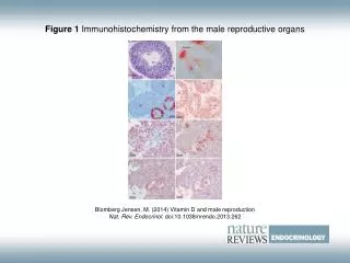
Applications • Cancer diagnostics • differential diagnosis • Treatment of cancer • Research
Part 1 Tissue preparation 1. Fixation Fresh unfixed, fixed, or formalin fixation and paraffin embedding 2. Sectioning 3.Whole Mount Preparation
pretreatment Part 2 1. Antigen retrieval Proteolytic enzyme method and Heat-induced method 2. Inhibition of endogenous tissue components 3% H2O2, 0.01% avidin 3. Blocking of nonspecific sites 10% normal serum
Part 3 staining • Make a selection based on the type of specimen, the primary antibody, the degree of sensitivity and the processing time required.
Controls • Positive Control It is to test for a protocol or procedure used. It will be ideal to use the tissue of known positive as a control. • Negative Control It is to test for the specificity of the antibody involved.