Structure
Structure. Brain Imaging Techniques Localization of Function Hemispheric Asymmetry. Brain Imaging Techniques. Can experience effect the way the brain functions? If you learned to no longer fear a situation, what would be the physiological effect?

Structure
E N D
Presentation Transcript
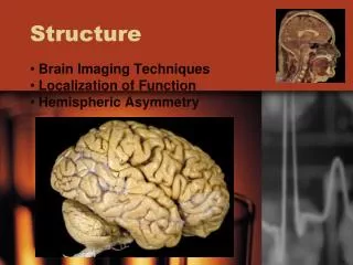
Structure Brain Imaging Techniques Localization of Function Hemispheric Asymmetry
Brain Imaging Techniques • Can experience effect the way the brain functions? • If you learned to no longer fear a situation, what would be the physiological effect? “There is no magic, it is just magic when we don’t know what is going on.”
Brain Imaging Techniques • Phineas P. Gage (1823-1860) • Railroad construction foreman who had a large iron rod driven completely through his head, destroying one or both of his brain’s frontal lobes. • The damage resulted in behavioral and personality changes that were very profound. • Phineas Gage influenced 19th-century thinking about the brain and localization of function. • First case suggesting that damage to specific regions of the brain might affect personality and behavior.
Brain Imaging Techniques EEG (electroencephalogram) • Recording of neural activity detected by electrodes. • Records brain noise. PET (positron-emission tomography) • Injecting radioactive isotopes. • Increased activity results in different colors. MRI(magnetic resonance imaging) • Radio frequencies are used. • The nuclei in atoms of cells turn differently and vibrate differently when hit with different waves. • Thus you can gear your scan towards specific cells in the body. CAT (computed axial tomography) • Uses tomography; where digital geometry processing is used to generate a three-dimensional image of the internals of an object from a large series of two-dimensional X-ray images taken around a single axis of rotation.
Brain Imaging Techniques EEG: MRI: PET:CAT:
Localization of Function Localization of function: Specific regions perform different (though overlapping) tasks. • Brain Imaging Techniques • Franz Joseph Gall • Developed Phrenology
Localization of Function Information processing in humans and animals operate similarly.
Localization of Function • Why would a dolphin have such a large brain? • What part of their brain would you expect to be more advanced than ours? • Less advanced?
Localization of Function • Hindbrain • Pons • Cerebellum • Medulla • Midbrain • Superior/Inferior Colliculi • Tectum • Aqueduct of Sylvius • Substantia Nigra • Reticular Activating System • Red Nucleus • Forebrain • Diencephalon • Telecencephalon
Localization of Function Cerebrum: Largest brain structure. • Controls: Sensory, Motor, Cognition • Division: Cerebral cortex, Lobes • Lateralization: Each half specializes. Corpus Callosum: • Connects the left and right hemisphere. • Allows the hemispheres to communicate. Commissurotomy: • Severing the corpus callosum. • Used with patients that have seizures.
Localization of Function Prefrontal Cortex Divisions: • Dorsal • Imitation • Planning • Logic • Sequencing • Ventral • Emotional Modulation
Localization of Function Prefrontal lobes structure our world for us and set the path for our way of thinking. Basically they regulate how we are going to appraise and approach any situation which affects everything from stress reactions to our immune response, to our working memory.
Localization of Function • Fusiform Gyrus: • Processing of color information • Face and body recognition • Word recognition • Number recognition • Within-category identification • Prosopagnosia: (face blindness) • Disorder of face perception where the ability to recognize faces is impaired, while the ability to recognize other objects may be relatively intact.
Localization of Function The ventricular system is a set of structures containing cerebrospinal fluid in the brain. It is continuous with the central canal of the spinal cord.
Localization of Function I. Brain Stem: • Medulla: • Bodily functions we don’t have to control. • Respiration and heart beat. • Pons: • Sleeping. • Waking. • Dreaming. • Reticular Activating System(RAS): • Distributes incoming stimulation throughout the brain. • Controls sleeping and waking. • Utilizes our Zeitgeber (Time-Giver).
Localization of Function II. Cerebellum: • Controls balance, smoothness of movement, starting movement, cessation of movement • Kinesthetic sense III. Thalamus: • Sends sensory messages to the cerebral cortex • Determines which higher processing center is best to interpret a given message. • Vision – Occipital Lobe. • Sound – Temporal Lobe. • Tactile – Postcentral Gyrus, Parietal Lobe. • Taste – Cerebral Cortex, Hypothalamus, Amygdala. • Smell – Does not pass through. Processed in the olfactory bulb, amygdala, and hypothalamus.
Localization of Function IV. Hypothalamus Hypothalamus: Involved in emotions and drives vital to survival: (autonomic nervous system) • Fear • Hunger • Thirst • Reproduction • Pituitary gland: Endocrine gland that releases hormones and regulates the other endocrine glands. • “Master Gland”
Localization of Function V. Amygdala: • Evaluates sensory information, determines importance. • Approach and withdraw reaction. • Accesses danger or threat to the body. • Dysfunction in this area causes: Depression • Controls genuine smile-Dewshane Smile • Major regions- • Central Nucleus- “Go” or “No-Go” • Medial Nucleus- Smell
Localization of Function VI. Hippocampus: • Storage of new information in memory. • Short term memory • If something is not successfully stored in short term memory then it will never make it into long term memory • Dreaming. • Rat Hippocampus (stained • green-Nissl)
Localization of Function VII. Limbic System: • Hypothalamus, Amygdala, Hippocampus • Involved in: • Emotional reactions. • Motivated behavior. • Located between the higher order and lower order processing centers of the brain.
Localization of Function • When behavior has adaptive properties the VTA releases Dopamine and sends it to the Nucleus Accumbens which causes LTP both there and in the Forebrain. • This increases the likelihood of repeating the behavior.
Localization of Function • Broca’s Area: Responsible for speech production. • Inferior Frontal Gyrus • Wernicke's Area: Responsible for language comprehension. • Posterior section of the Superior Temporal Gyrus.
Sex and Neuroscience • Structures that are larger in the healthy female brain, relative to cerebrum size • SA (sexual arousal) evoked in females produced a non-significant hypothalmic activation. Women respond less to erotic viewing films... • Structures that are larger in the healthy male brain, relative to cerebrum size • Men experience a greater SA to visual erotic stimuli than women. This also produced a more intense activation of the hypothalmic area