Download

1 / 7
90 likes | 375 Vues
Monoclonal Antibody Production. Cathy Langford, Judy Knadler, Jamie Wolf. Monoclonal Antibody Production Method.

E N D
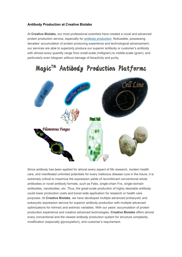
Monoclonal Antibody Production Cathy Langford, Judy Knadler, Jamie Wolf
Monoclonal Antibody ProductionMethod • Monoclonal Antibody Production technology was developed in 1975. Since its development it has been very important in the modern medical science with the diagnosis, therapy, research and even basic science today. It is still largely dependent upon animal testing however. Because it requires immunization of mice in order for them to create the antibodies to be grown. • Monoclonal Antibody Production or mAb is produced by cell lines or clones obtained from the immunized animals with the substance to be studied. Cell lines are produced by fusing B cells from the immunized animal with myeloma cells. To produce the desired mAb, the cells must be grown in either of two ways: by injection into the peritoneal cavity of a suitably prepared mouse (the in vivo, or mouse ascites, method) or by in vitro tissue culture. • The vitro tissue culture is the method used when the cells are places in culture outside the mouse's body in a flask.
Explanation Of Picture • The previous picture is showing a mouse being immunized against a target cell “X”. This will allow the mouse to produce antibodies for that will target against the “X” antigen. • Once the mouse has formed antibodies to the “X” antigen the cells are then isolated in the mouse’s spleen. Monoclonal antibodies are produced by fusing single antibody-forming cells to tumor cells grown in culture. The resulting cell is called a hybridoma. Hybridoma cells are continuously growing cell line generated by the fusion of a myeloma cell and a normal cell that are capable of producing antibodies. • Each hybridoma will produce relatively large quantities of identical antibody molecules. Because the hybridoma is multiplying in culture, it is possible to produce a population of cells, each is producing identical antibody molecules. These antibodies are called "monoclonal antibodies" because they are produced by the identical offspring of a single, cloned antibody producing cell.
Why this method is used!! • This method is used because antibodies must be formed from the immunization of the substance being studied. So antibodies must be produced. Once the antibodies are produced the animal aspect of the study can be eliminated and tissue culture can then be used. • When using live mice researchers have found that it is the better option because in vitro doesn’t always produce adequate cell lines that are adaptive to tissue culture. Protein denaturation can occur from purification techniques and antibody activity is decreased with normal activity not represented. Also cell lines could possibly become contaminated when using in vitro technique.
Monoclonal ELISA test system run. Labels across the top of the plate indicate the capture antibody used in the designated column: P, polyclonal rabbit anti-Lp 1 through 9, the mouse monoclonal antibody indicated by the same number; M, polyclonal normal mouse immunoglobulin. Labels on the side of the plate indicate the specimen tested in that row: A, PBS-BSA; B through D, positive lung homogenate extracts from cases in the Philadelphia outbreak; E, Philadelphia 1 culture extract; F, OLDA culture extract; G, Knoxville 1 culture extract; H, negative lung homogenate extract.
References • http://www.accessexcellence.org/RC/VL/GG/monoclonal.html • http://www.med.nyu.edu/dlar/iacuc/monoclonal.html • http://orsted.nap.edu/openbook.php?record_id=9450&page=5 • SUSAN L. BROWN,* WILLIAM F. BIBB, AND ROGER M. McKINNEY Use of Monoclonal Antibodies in an Epidemiological MarkerSystem: a Retrospective Study of Lung Specimens from the 1976Outbreak of Legionnaires Disease in Philadelphia by IndirectFluorescent-Antibody and Enzyme-Linked Immunosorbent; AssayMethodsJOURNAL OF CLINICAL MICRODIOLOGY, Jan. 1985, p. 15-19