Download
1 / 69
710 likes | 1.07k Vues
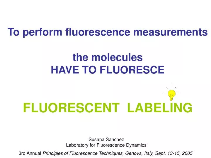
To perform fluorescence measurements the molecules HAVE TO FLUORESCE FLUORESCENT LABELING. Susana Sanchez Laboratory for Fluorescence Dynamics. 3rd Annual Principles of Fluorescence Techniques, Genova, Italy, Sept. 13-15, 2005. How to choose the labeling protocol?

E N D
To perform fluorescence measurements the molecules HAVE TO FLUORESCE FLUORESCENT LABELING Susana Sanchez Laboratory for Fluorescence Dynamics 3rd Annual Principles of Fluorescence Techniques, Genova, Italy, Sept. 13-15, 2005
How to choose the labeling protocol? Light source available In vivo or in vitro Lifetime of the fluorescent probe Spectroscopy or Microscopy
Can the wrong labeling induce errors in interpretation? Experimental considerations
Illumination Volume and sample concentration FCS One or two-photon Spectroscopy 0.4x0.4x1cm 0.16 cm3 (160uL=160x10-6L) 1x10-15 L Number of molecules in the excitation volume 1 uM 9.7x1013603 10 nM 9.7x10116
Correct labeling for the chosen technique Example: dimer dissociation Spectroscopy: Polarization measurements Measuring a population of molecules Microscopy: FCS measurements Number of molecule change 2 D dimer/D monomer 1.2 Measuring single molecules level Number of molecule change 1
Dcoef log Too many labeled particles
Giant Unilamellar Vesicles Large Unilamellar Vesicles 50nm a 400nm (0.05-0.4 mm) 10-100mm 80 um Measuring a population of liposomes Measuring single liposomes Observing populations or particular behavior
Labeling proteins Labeling DNA Labeling membranes Coffee break Quantum dots Ions indicators Labeling “in vivo”
Naturally Occurring Fluorophores in Proteins aromatic amino acids Phenylalanine (Phe – F) Ex/Em 260 nm/282 nm Tyrosine (Tyr – Y) ex/em 280 nm/305 nm Tryptophan (Trp-W) ex/em 280, 295nm/ 305-350 nm
NADH (oxido-reductases) Ex/Em 340/460 nm FAD (metabolic enzymes (ex/em 450nm/540 nm) Porphyrins (ex/em 550 nm/620 nm), Enzymes Cofactors
quantum yield similar to that of tryptophan . • small and solvent-insensitive Stokes shift 5-Hydroxytryptophan ex/em 310nm/339 nm • low quantum yield and a large Stokes shift in water 7-azatryptophan ex/em 320nm/403nm Protein Science (1997), 6, 689-697. Synthetic Fluorophores in Proteins Tryptophan derivatives Advantage: absorbance spectrum of these analogues is red-shifted with respect to that of tryptophan. Therefore it is possible to selectively excite them, in proteins, in the presence of tryptophan of other proteins or DNA bases.
cyanobacteria red algae Fluorescent proteins Phycobiliproteins • -Intensely fluorescent proteins from red algae and cyanobacteria (blue-green algae). • -Absorb strongly between 470 and 650 nm. • - In intact phycobilisomes, they are only weakly fluorescent, due to efficient energy transfer to photosynthetic reaction centers. Highly fluorescent in vitro. Four main classes of phycobiliproteins.
Green Fluorescent Protein (GFP) - from the bioluminescent jellyfish Aequorea victoria. - Obvious -barrel structure, with chromophore housed within the barrel. - Remarkably, the chromophore is formed spontaneously (from Ser-65, Tyr-66, Gly-67) upon folding of the polypeptide chain, without the need for enzymatic synthesis. - As a result, it is possible to insert the gene for GFP into cells and use the resulting protein as a reporter for a variety of applications.
Extrinsic probes (not present in the natural molecule/macromolecule) Non-covalent Attachments Ethidium bromide Interact with dsDNA & IgG/Antigen bis-ANS binds to hydrophobic patches on proteins Mant-GDP
Fluorescent group Reactive group Reactive group aa Covalent Attachments Available reactive group in the protein Light source Lifetime of the fluorescent group Spectral properties Autofluorescence Labeling should not change the biological activity of the protein.
Fluorescent group Reactive group Reactive group aa BODIPI (493/503), t= FITC (488/512) t=4.05 Dansyl chloride IAEDANS (360/480) t =15 ns Pyrenebutyric acid Texas Red ANS (374/454) t (EtOH)= 8 ns NBD
Fluorescent group Reactive group + Reactive group aa Targeting amino groups + Lysine Arginine
Fluorescent group Reactive group + Reactive group aa Targeting thiol groups: + Cysteine
Removal of the free dye Protein in buffer Incubation time Addition of the fluorescent dye Biological testing Sample characterization Labeling ratio Absorption spectra Protein determination Activity measurements SDS or native gel Denaturation exp etc. [protein] [fluorescent dye] General labeling protocol for extrinsic labeling
Labeling DNA http://info.med.yale.edu/genetics/ward/tavi/n_coupling.html
5’ 3’ A B 3’ 5’ 5’ 3’ 3’ 5’ Nick translation DNase nicks the double stranded DNA. E.Coli Pol I has 5’-3’ exonuclease activity has 5’-3’ polymerizing activity End labeling of fragments dUTP 200-500 bp DNase I, which in the presence of Mg++ ions becomes a single stranded endonuclease creates random nicks in the two strands of any DNA molecule. E. coli polymerase I, it's 5'-3' exonuclease activity removes nucleotides "in front" of itself. the 5'-3' polymerase activity adds nucleotides to all the available 3' ends created by the DNase . This exonuclease/polymerase activity, moves (or "translates") any single stranded nick in the 5'-3' direction. When nicks on opposite strands meet, the DNA molecule breaks
5’ 5’ 3’ A B C 3’ 5’ 5’ dUTP 5’ 3’ 5’ 5’ 5’ 5’ PCR Taq Pol incorporates nucleotides along the entire length of the DNA. Higher labeling efficiency by PCR. Requires decreased amount of probe. 100-5,000 bp Two single stranded DNA primers (18-30 bp long), one forward and one reverse are synthesized (yellow arrows). After adding the primers, the Taq polymerase, the reaction mix is denatured Then, the primers are allow to anneal (Fig. 2a) to their target sequences (annealing step). Then Taq polymerase synthesize the new DNA strands (extension step, Fig 2b).
Labeled dUTP labeled nucleotides are synthesized by chemically coupling allylamine-dUTP to succinimidyl-ester derivatives of 1- fluorescent dyes 2- haptenes (Biotin, Digoxigenin, Dinitrophenyl - these require fluorescently-labeled antibodies or specific proteins for visualization/detection). Commercially labeled dUTP fluorescein-aha-dUTP from Molecular Probes
Fatty acids analogs and phospholipids Sphingolipids, sterols,Triacylglycerols etc. Dialkylcarbocyanine and Dialkylaminostyryl probes. Other nonpolar and amphiphilic probes. Laurdan, Prodan, Bis ANS
DPH (D202), cis-parinaric acid (P36005), BODIPY 500/510 C4 NBD-C6-HPC (N3786), N-Rh-PE (L1392), bis-pyrene-PC (B3782), DiA (D3883) DiI (D282), C12-fluorescein (D109). Membrane probes
Liquid crystalline phase Gel phase Laurdan Weber, G. and Farris, F. J.Biochemistry, 18, 3075-3078 (1979) .
Ex=340 nm IB IR -0.2 loose lipid packing 0.6 tight lipid packing Laurdan Generalized Polarization (GP) Parasassi, T., G. De Stasio, G. Ravagnan, R. M. Rusch and E. Gratton. Biophysical J., 60, 179-189 (1991).
DPPC DPPS:DPPC (2:1) DPPG:DPPC (2:1) DMPA:DMPC (2:1) DPPG:DLPC (1:1) DPPC:DLPC (1:1) + GP Temperature (°C) GP in the cuvette MLVs,SUVs,LUVs Lipid Phase Transition Parassassi, Stasio, Ravaganan, Rusch, & Gratton (1991) Biophys. J. 60, 179
GP in the microscope (2-photon) Measurement of Laurdan in the GUVs using SimFCS software ch1 Blue filter ch2 Red filter GP image GP histogram
31% chol 0%chol 25 37 25 37 C T C T Temperature (Celsius) -1 GP 1 Temperature (Celsius)
DOPC/DPPC 1:1mol/mol (31% mol chol) 24.8°C, rHDL 96Å
core composed of cadmium sulfide (CdS), cadmium selenide (CdSe), or cadmium telluride (CdTe). The semiconductor material is chosen based upon the emission wavelength, however it is the size of the particles that tunes the emission wavelength . shell biomolecule In the cores emission is typically weak and always unstable. 1-Reorganization of crystalline imperfections and defects results in sites known as traps. These sites provide a non-productive, non-emissive, pathway. 2- The re-organized surfaces tend to be very reactive and they easily become polluted by solvent molecules, air molecules, impurities, etc., The shell material (typically ZnS in Qdots) has been selected to be almost entirely unreactive and nearly completely insulating for the core. coating a layer of organic ligands covalently attached to the surface of the shell which further passivates the core-shell and acts as a glue to the outer layer. the outer layer is a mixed hydrophobic/philic polymer. The hydrophobic part interacts with the inner coating while the hydrophilic portion interacts with the external solvent to provide solubility in buffers. This coating provides a flexible carboxylate surface to which many biological and nonbiological moieties can be attached. The resulting surface is derivatizable with antibodies, Streptavidin, lectins, nucleic acids, and related molecules of biological interest.
fuorescein Typical water-soluble nanocrystal (NC) sample in PBS Their emission spectra is narrow and symmetrical. The emission is tunable according to their size and material composition, allowing closer spacing of different probes without substantial spectral overlap. They exhibit excellent photo-stability. They display broad absorption spectra, making it possible to excite all colors of QDs simultaneously with a single excitation light source and to minimize sample autofluorescence by choosing an appropriate excitation wavelength.
The emission is tunable according to their size and material composition Samples were placed in front of a common UV hand lamp. All samples are induced to emit their respective colors even though a single source was used to excite them. The colored spheres illustrate the relative sizes of the CdSe quantum dots in the vials.
Example Wu et al. Nature Biotechnology21, 41 - 46 (2002) • Microtubules were labeled with • 1-monoclonal anti-tubulin antibody. • 2- biotinylated anti-mouse IgG and QD 630−streptavidin (red). • (B) Control for (A) without primary antibody. • (C) Actin filaments were stained with • 1-biotinylated phalloidin and QD 535−streptavidin (green). • (D) Control for (C) without biotin-phalloidin. • The nuclei were counterstained with Hoechst 33342 blue dye.
Cations: H+, Ca2+, Li+, Na+, K+, Mg2+, Zn2+, Pb2+and others Anions: Cl-, PO42-,Citrates, ATP, and others Fluorescent probes for Ions Fluorescence probes have been developed for a wide range of ions:
How do we choose the correct probe for ion determination? 1-Dissociation constant (Kd) Must be compatible with the concentration range of interest. The Kd of the probe is dependent on pH, temperature, viscosity, ionic strength etc. Calibration is important. 2- Measurement mode Qualitative or quantitative measurements. Ratiometric measurements. Illumination source available. 3- Indicator form (salt, Cell-permeant acetoxymethyl estes or dextran conjugate) Cell loading and distribution of the probe. Salt and dextran…microinjection, electroporation, patch pipette. AM-esters ….cleaved by intracellular esterases
Probes For Calcium determination UV FURA ( Fura-2, Fura-4F, Fura-5F, Fura-6F, Fura-FF INDO ( Indo-1, Indo 5F) VISIBLE FLUO (Fluo-3, Fluo-4, Fluo5F, Fluo-5N, Fluo-4N) RHOD ( Rhod-2, Rhod-FF, Rhod-5N) CALCIUM GREEN (CG-1, CG-5N,CG-2) OREGON GREEN 488-BAPTA (OgB-1, OgB-6F, OgB-5N, OgB-2) FURA
FURA-2 Ratiometric: 2 excitation /1emission
Indo-1 Ratiometric: 1excitation /2emission
Calcium Green-1 Calcium Green-2
Probes For pH determination Table 20.1 — Molecular Probes' pH indicator families, in order of decreasing pKa
BCECF In situ calibration can be performed by using the ionophore nigericin (N1495) at a concentration of 10~50 μM in the presence of 100~150 mM potassium to equilibrate the intracellular pH with the controlled extra cellular medium
Example 1 K.Hanson, M.J.Behne, N.P.Barry, T.M.Mauro, E.Gratton. Biophysical Journal. 83:1682-1690. 2002. Dye in DMSO is applied to the a live animal and incubated for some time Labeled skin is removed imaging