Download
1 / 13
140 likes | 512 Vues
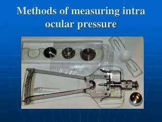
A Wireless, Implantable Intra-Ocular Pressure Sensor for the Management of Glaucoma. Gabriel Simon, M.D. Ph.D. Sept 16, 2008 ESCRS. Current IOP Measurements. Current Methods of IOP Measurement Applanation Tonometer (Goldmann) Requires contact with eye surface

E N D
A Wireless, Implantable Intra-Ocular Pressure Sensor for the Management of Glaucoma Gabriel Simon, M.D. Ph.D. Sept 16, 2008 ESCRS
Current IOP Measurements • Current Methods of IOP Measurement • Applanation Tonometer (Goldmann) • Requires contact with eye surface • Errors due to corneal thickness, past surgeries, etc.. • Clinical & limited home use • Dynamic Contour Tonometry (PASCAL DCT) • Relies on contour matching • More accurate than applanation tonometers • Clinical use only • Electronic Indentation Tonometer (Tono-Pen) • Limited accuracy • Home and clinical use • No method for continuous, remote IOP monitoring currently exists • Continuous monitoring will allow complete glaucoma management • RF Transmission of Data & Power • Current range exceeds 3 meters
Background – IOP Monitoring • Current Approach – Reactive • Sample every 3-6 months • Tonometer measurement, clinic-based • Corneal thickness-induced errors • IOP Fluctuations can vary over 24-hour period • Diurnal variations in IOP (2008 Sit, et al.) • Circadian/Hourly fluctuations (2006 Barkana, et al.) • Continuous IOP Monitoring – Managing IOP • Sample continuously, with daily upload • Accurate to 0.5 mmHg • Ophthamologist can monitor IOP trends daily • Requires: • Wireless, high-sensitivity sensor • Ultra-low power circuitry • Miniature packaging (<5mm per side) for implantation • External RF-charging and data collection device
Implantable Sensor Concept • Implantable sensor for continuous patient monitoring • Prototyped at Purdue Brain-Computer Interface (BCI) Lab • Implanted in anterior chamber orsuprachoroidal space • 300µm overall thickness • Capacitive Sensor • 0-50mmHg sensitivity • 0.5mmHg accuracy • Amplifier and Telemetry • Capture IOP every 5 minutes • RF download and recharge • External Unit • Stores & transmits all IOP data • Held to eye for <10 seconds • Provides significant improvement in quality of care
IOP Sensor Device • Remote patient monitoring • Provides near-continuous IOP data on daily basis • Disease progression & drug monitoring • Continuously sampled IOP data evaluated daily • Email or internet interface, Bluetooth compatible • E-consultation if necessary • Minimize cost & time while improving quality of care • Daily reports of IOP vs. sporadic visits • Patient and Doctor Interface • Promotes patient compliance • Easy to use for physicians • IOP trends and warnings based on relevant information
Amplifying module Wireless data module antenna mixer LNA LPF amp S2 S1 VCO Clk Biasing circuit External receiver with graphical user interface and power-coupling hardware Voltage regulator Battery 1) Amplifying module 2) Wireless data module 3) IOP sensing module 4) Powering module 5) External user interface IOP sensing module Powering module Intra-Ocular Pressure Sensor Design Research partners: Gabriel Simon2, Babak Ziaie3, SOLX4 2Professor of Ophthalmology, Boston University 3Associate Professor, Department of Electrical Engineering, Purdue University 4Boston University startup company
IOP Sensor Concept – Location • Suprachoroidal Implant • Sensor surface protrudes into Anterior Chamber • IOP measured in AC • 15-minute implant procedure • Posterior Chamber: same as IOL procedure • Similar to gold shunt/suprachoroidal procedure • Minimal, transient complications • Posterior Chamber Implant • Same as IOL procedure • No learning curve • 5-10 minute procedure • Candidate patients • Glaucoma patients • Cataract - IOL patients • Does not preclude later IOL implant
Implant Prototypes and Materials • Implant materials: • Low-TemperatureCo-fired Ceramic (LTCC) • Silicon • PMMA • Liquid-Crystal Polymer (LCP) Silicon substrate LTTC substrate
Posterior Chamber Sensor Implant PMMA or Liquid-Crystal Polymer (LCP) substrate
Wireless Power Transmission • Initial studies show that we can achieve low-power RF transmission from a miniature implantable device for ocular implant applications. • In-vivo experiments show that the implant was measured to have a sufficient signal-to-noise ratio margin for high data-rate transmission, validating this approach for intra-ocular pressure telemetry.
Wireless Transmission - Testing • The power received is at least 10 dB greater than the MDS (minimum detectable signal) and we can achieve successful wireless data transfer. • LTCC based loop antennas provide less attenuation caused by tissues after implanting than the silicon based monopole antennas.
IOP Sensor Data Management • Data Collection • Patient recharges & uploads once/day • Data Hub at patient’s residence • Relies on PC with modem, phone line, or cell phone • Data Management • Multi-user, web-based database server • HIPAA compliant, with backup security • Allows multi-point access via internet • Clinical Data Analysis • Patient information provided to clinician via internet UI • IOP summary trends • Medication compliance • Capable of supporting additional physiological data capture • Presented to clinician in simple overview • 1-5 minutes of review per patient • 24-7 access to database with subscription
Aqueous Outflow Summary and Future Studies • Continuous, wireless IOP sensor will provide a new level of glaucoma management • Remote patient monitoring via internet • Improved treatment, compliance, and outcomes • Reduced office visits and cost-to-treat • Future: Combination diagnostic and treatment • Passive suprachoroidal sensor with shunting capabilities • flow channels incorporated into sensor package • Active suprachoroidal glaucoma manage-ment device • Remote IOP monitoring capability • Adjustable flow resistance • Wireless adjustment of outflow facility • Based on IOP signal from sensor • Remote adjustment via external charging device