Jaffe GJ, Martin DF, Toth CA, Daniel E, Maguire, MG,
130 likes | 288 Vues
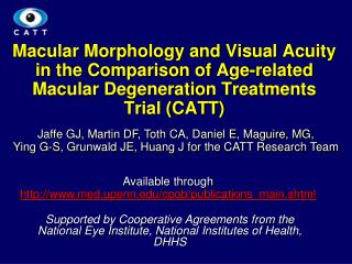
Macular Morphology and Visual Acuity in the Comparison of Age-related Macular Degeneration Treatments Trial (CATT ). Jaffe GJ, Martin DF, Toth CA, Daniel E, Maguire, MG, Ying G-S, Grunwald JE, Huang J for the CATT Research Team.

Jaffe GJ, Martin DF, Toth CA, Daniel E, Maguire, MG,
E N D
Presentation Transcript
Macular Morphology and Visual Acuity in the Comparison of Age-related Macular Degeneration Treatments Trial (CATT) Jaffe GJ, Martin DF, Toth CA, Daniel E, Maguire, MG, Ying G-S, Grunwald JE, Huang J for the CATT Research Team Available through http://www.med.upenn.edu/cpob/publications_main.shtml Supported by Cooperative Agreements from the National Eye Institute, National Institutes of Health, DHHS 1
Presence and Type of Fluid on OCT Over Time Jaffe et al. Macular Morphology and Visual Acuity in the Comparison of Age-related Macular Degeneration Treatments Trials Ophthalmology Epub 2013 May 03
Impact Over Time of Drug & Dosing Regimen on OCT–Determined Thickness Measurements Jaffe et al. Macular Morphology and Visual Acuity in the Comparison of Age-related Macular Degeneration Treatments Trials Ophthalmology Epub 2013 May 03
Retinal thickness category over time by treatment group Jaffe et al. Macular Morphology and Visual Acuity in the Comparison of Age-related Macular Degeneration Treatments Trials Ophthalmology Epub 2013 May 03
Lesion Components Under the Foveal Center by Drug and Dosing Regimen Jaffe et al. Macular Morphology and Visual Acuity in the Comparison of Age-related Macular Degeneration Treatments Trials Ophthalmology Epub 2013 May 03
Foveal Lesion Composition Baseline Week 52 Jaffe et al. Macular Morphology and Visual Acuity in the Comparison of Age-related Macular Degeneration Treatments Trials Ophthalmology Epub 2013 May 03
Ranibizumab Monthly (A) (B) Bevacizumab Monthly Geographic Atrophy Geographic Non - geographic 2.6% Atrophy 0.8% Fluid Only 8.2% Non - geographic Atrophy Atrophy 11.8% 18.5% Fluid Only 10.9% Blocked Blocked CNV 21.2% Fluorescence Fluorescence CNV 4.9% 5.3% 21.5% Can't Grade 1.6% Can't Grade SPED 0.4% SPED 3.0% No Pathology 0.4% 24.9% Scar 20.4% No Pathology Scar 17.7% RPE Tears 0.8% RPE Tears Other 16.2% Other 4.9% 1.5% 2.3% (C) Ranibizumab PRN (D) Bevacizumab PRN Geographic Non - geographic Geographic Atrophy 1.1% Fluid Only 9.4% Atrophy 14.5% Atrophy 3.1% Non - geographic Fluid Only 4.5% Atrophy 13.3% CNV 29.8% Blocked Blocked Fluorescence Fluorescence CNV 26.5% 3.4% 1.6% Can't Grade Can't Grade 3.4% 2.4% No Pathology No Pathology SPED 1.1% 16.1% 20.1% SPED 0.4% Other 3.9% Scar 18.8% Scar 19.0% Other 4.8% Hemorrhage Hemorrhage RPE Tears 0.8% 0.4% 0.7% RPE Tear 0.7% Involvement of the foveal center by CNV or sequelae of CNV at week 52
Mean visual acuity by status of fluid Jaffe et al. Macular Morphology and Visual Acuity in the Comparison of Age-related Macular Degeneration Treatments Trials Ophthalmology Epub 2013 May 03
Retinal thickness and Visual Acuity at Baseline and Follow-up Jaffe et al. Macular Morphology and Visual Acuity in the Comparison of Age-related Macular Degeneration Treatments Trials Ophthalmology Epub 2013 May 03
Nonlinear relationship of visual acuity with foveal total thickness during follow-up Jaffe et al. Macular Morphology and Visual Acuity in the Comparison of Age-related Macular Degeneration Treatments Trials Ophthalmology Epub 2013 May 03
Mean VA by Neovascular Lesion Area and Pathology in Foveal Center at Week 52 (N=1053) *1-way analysis of variance RPE = retinal pigment epithelium §Other includes pigment, drusenoid pigment epithelial detachment and non-leaking choroidal neovascularization Jaffe et al. Macular Morphology and Visual Acuity in the Comparison of Age-related Macular Degeneration Treatments Trials Ophthalmology Epub 2013 May 03
Adjusted Mean Visual Acuity for OCT and Fundus Features at Week 52 (n=1004)∗ *Subjects (n=49) with missing data for fluid or retinal thickness were excluded. CNV= choroidal neovascularization RPE = retinal pigment epithelium Jaffe et al. Macular Morphology and Visual Acuity in the Comparison of Age-related Macular Degeneration Treatments Trials Ophthalmology Epub 2013 May 03
Conclusions Jaffe et al. Macular Morphology and Visual Acuity in the Comparison of Age-related Macular Degeneration Treatments Trials Ophthalmology Epub 2013 May 03 • Anti–vascular endothelial growth factor (VEGF) therapy reduced lesion activity and improved VA in all treatment groups. • At all time points, eyes with residual IRF had worse VA than those without. • Eyes with abnormally thin or thick retinas, residual large lesions, and scar also had worse VA. • Monthly ranibizumab dosing yielded more eyes with no fluid and an abnormally thin retina, although the long-term significance is unknown. • These results have important treatment implications in eyes undergoing anti-VEGF therapy for neovascular