Download
1 / 24
350 likes | 1.55k Vues
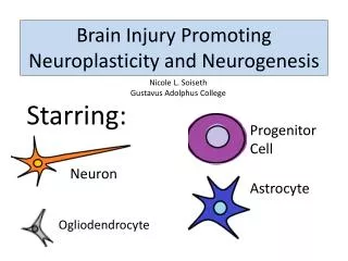
Neuroplasticity and Rehabilitation Strategies. Robert K. Shin M.D. VA MS Center of Excellence Assistant Professor Departments of Neurology and Ophthalmology University of Maryland School of Medicine. Neuroplasticity?. The ability of cortex to reorganize in response to injury. Question.

E N D
Neuroplasticity and Rehabilitation Strategies Robert K. Shin M.D. VA MS Center of Excellence Assistant Professor Departments of Neurology and Ophthalmology University of Maryland School of Medicine
Neuroplasticity? • The ability of cortex to reorganize in response to injury
Question • Is the brain compensating for damage from MS in order to maintain normal function?
Functional MRI • BOLD contrast analysis • Oxyhemoglobin • Deoxyhemoglobin • Alternating periods of task vs. rest • Activated regions determined statistically
A dynamic cortical response? • Initially increased activation of contralateral sensorimotor cortex • Ipsilateral activation seen initially as well • Activation gradually reduced as patient recovered
Clinically isolated syndrome • 16 consecutive patients with a CIS suggestive of multiple sclerosis • 15 age- and sex-matched controls • Functional MRI during finger flexion Rocca et al. NeuroImage 2003;18:847-855
A response to cortical pathology? • Decreased NAA found in clinically stable CIS patients • Increase in activation of somatomotor cortex correlated with worsening brain damage
Another puzzle • Optic neuritis causes vision loss and prolonged VEPs • Vision in optic neuritis almost always recovers • VEPs frequently remain abnormal
Question • Is the brain somehow compensating for impaired optic nerve function?
Recovery from optic neurits • 7 patients who had recovered from optic neuritis • 7 controls • Functional MRI during photic stimulation Werring, et al. JNNP 2000;68:441-449
Functional reorganization? • Only occipital activation seen in controls • Additional extra-occipital areas were activated in patients who had recovered from optic neuritis
Attention and memory • 22 patients with RR-MS • 22 age-matched controls • Functional MRI study • Paced Auditory Serial Addition Test (PASAT) • Recall task Mainero, et al. NeuroImage 2004;21:858-867
PASAT Mainero, et al. NeuroImage 2004;21:858-867
Recall Mainero, et al. NeuroImage 2004;21:858-867
T2 LL Mainero, et al. NeuroImage 2004;21:858-867
An adaptive mechanism? • Altered activation during cognitive tasks in MS patients • Activation increases as T2 lesion load increases • But activation is greater in patients with better function
Conclusions • Functional MRI activity is altered in MS patients • These changes appear to be an adaptive response to brain damage
Implications for rehabilitation? • Can functional MRI be used to prognosticate? • Can “cortical plasticity” be enhanced? • Proprioceptive stimulation • Forced use • Neurotrophic factors • Increased neurotransmitter release